Hedgehog worms
| Hedgehog worms | ||||||||||||
|---|---|---|---|---|---|---|---|---|---|---|---|---|

|
||||||||||||
| Systematics | ||||||||||||
|
||||||||||||
| Scientific name | ||||||||||||
| Echiura | ||||||||||||
| Newby , 1940 | ||||||||||||
| Orders | ||||||||||||
|
Types (selection) |
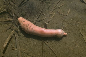
The hedgehog worms (Echiura or Echiurida) are a class of annelids (Annelida) with around 150 known species . These exclusively marine animals inhabit various marine habitats around the world, from the intertidal zone to deep- sea trenches up to a depth of 10,000 meters . Hedgehog worms inhabit hemisessil soft soils, less often crevices and caves in hard substrates.
morphology
Morphology of the adult animals
The size of hedgehog worms is diverse, ranging from a few decimeters to several meters.
The unsegmented body can be divided into the anterior, preoral section, the prostomium (proboscis = trunk), and the posterior section, the trunk. The prostomium exceeds the sack-shaped or cylindrical trunk many times over in length. The largest species, Ikeda taenioides , reaches lengths of up to 2 meters and only 40 centimeters make up the trunk. Despite its mobility and muscularity, the prostomium cannot be drawn into the trunk.
The epidermis, which is not ciliated except for the ventral side of the prostomium, is covered by a cuticle . A characteristic feature of the epidermis of hedgehogworms is a pair of bristles on the ventral side of the trunk, movable by muscles . Some species also have one or two rings of anal bristles. The fine structure of the cuticle and bristles is identical to that of the annelid worms. The epidermis forms numerous papillae in the trunk region, which give the impression that the animals are curled. The entire surface of the body is covered with serous gland cells.
Under the epidermis lies the powerful extracellular matrix (tissue part) in which gland cells, pigment cells, nerve cells and collagen fibers ( structural protein of the matrix) are embedded. The prostomium is almost completely filled with this connective tissue (cutis), where it also contains the complex muscles of the prostomium.
The muscles of the trunk lie under the matrix and consist of 8 layers of longitudinal, circular and diagonal muscles. The trunk also contains a Coelom cave (secondary body cavity), which is delimited by a peritoneum (serous peritoneum, lines the abdomen) and is only subdivided by atrophied mesenteries (fold in Coelom wall). Most species have an incomplete diaphragm in front of a pair of bristles . In front of this is a small space from which canals run into the prostomium . The coelom fluid consists of different types of coelomocytes.
The simple, closed blood vessel system consists of the ventral vessel, a dorsal vessel only formed in the front , the intestinal blood sinus and three prostomial vessels. The blood is colorless.
The uptake of oxygen takes place over the entire extent of the epidermis, in some species the rectum also serves to exchange gas. Water is constantly being pumped in and expelled again.
The anterior part of the trunk contains 1-2 pairs of metanephridia , which take up mature gametes (germ cells) from the coelomic fluid. These are then stored in a sack-shaped section.
The excretion takes place by numerous Analschläuche, which are provided with hoppers eyelashes and open into the rectum.
The intestinal canal can be divided into foregut, middle and rectum. There is a mouth opening at the base of the prostomium. From there the foregut leads via the pharynx , esophagus and perhaps also a goiter to the heavily convoluted midgut. He has a secondary intestine and an eyelash groove in the middle section.
The nervous system consists of a throat ring and an unpaired abdominal marrow . The former extends to the tip of the prostomium. There are ring-shaped branches on the abdominal medulla. In the prostomium, the two branches of the throat ring are connected by nerves.
Igelwümer do not have complex sensory organs, but a number of epidermal sensory cells are grouped into sensory papillae. A particularly high density was found on the prostomium.
The gonads are unpaired and are located on the ventral mesentery in the back of the trunk. The gametogenesis runs in the coelom. The mature sperm and egg cells are stored in the sac-shaped sections of the nephridia .
Most species do not show sexual dimorphism , but Bonellia viridis shows extreme differences in size between the sexes. Females sometimes reach torso lengths of 30 centimeters, while the male is only 2 millimeters long. For a long time, the males were thought to be parasitic platelets due to their differing morphology . This assumption was also confirmed at the time by the fact that the males live in the uterus of the females and fertilize the egg cells there. The sex determination here apparently takes place via a pheromone produced by the female , which causes the undifferentiated larvae to become dwarf males.
Morphology of the larval stage
The free-floating Trochophora larvae consist of epithelial and Hypo sphere . These are separated by the prototroch . The bottom of the upper or the top of the lower segment is horizontal. Characteristic of the larvae of hedgehog worms are other cilia , ocelles , a pair of protonephridia , an intestinal canal and mesoderm strips formed from mesoteloblasts . The abdominal marrow has its origin in a paired system of serially arranged cell groups.
Way of life
Hedgehog worms are hemisessile and rarely change their location in soft substrate; it is rarely hard substrate. The animals build a den in the substrate in which they spend almost their entire life. Depending on the species, the burrows have different shapes, with Echiurus echiurus it is U-shaped.
For food intake, only the prostomium is stretched out of the living corridor and passed over the substrate, but only the non-lashed dorsal side. The small food particles, preferably detritus and microorganisms , are brought to the cilia or muscles on the ciliate side, sorted and transported through the median eyelash groove to the mouth opening or later released again. This behavior leaves star-shaped feeding marks on the substrate surface.
All four species of the genus Urechis practice a special way of eating . With the help of prostomial glands, they build a mucous network in their living quarters. The animals can pump movements under this network; As a result, food particles are sucked in, which then get stuck in the net. If there is a need for food, the net owner eats the net with the food particles.
Reproduction
Hedgehog worms are always reproduced sexually; all species are separate sexes. Mostly, fertilization takes place in free water and outside the body of the parent animals. The eggs have a spiral groove . The young animals lead a planktonic way of life and sink to the ground after a maximum of 3 months. There they undergo a gradual metamorphosis in which the episphere becomes the prostomium and the hyposphere becomes the trunk.
Phenotypic sex fixation has been demonstrated in the species Bonellia viridis ; this species is a classic example of this in zoology. It is not factors that make the difference during and immediately after fertilization, but those after hatching. This peculiarity has not yet been fully clarified. If the larvae are out of contact with females for a long time, 78% develop into females and 1.5–3% into males, the rest become intersexes or die. This is different if they attach themselves to the prostomium of a female for four days: Then 75% develop into males and 15% into females, the others become intersex or die. In this way, numerous males can gather on one female; on one female there were already 85 males.
Systematics
According to the current state of knowledge , the hedgehog worms comprise around 150 species , which are divided into three orders :
The hedgehog worms were listed as an independent taxon in the rank of a tribe for a long time before they were assigned to the annelid worms (Annelida). They share numerous common features with them:
- The spiral furrowing of the eggs and the trochophora larvae
- Ultra structure of the bristles
- Fine structure of the cuticle
- Paired structure of the nervous system
- Formation of the mesoderm
- Morphology of the vascular system
The Vielborstern they agree with the intestinal canal ventral eyelashes channel and one of them separate by-intestinal under construction. The transformation of the prostomium into an organ that is used for food intake is an autapomorphy of hedgehog worms.
So far no efforts have been made to build a phylogenetic system within the hedgehog worms.
Hedgehog worms and humans
Since, due to the way of life of some species in the deep sea, there are no precise data on their populations, the level of threat to these animals is not known. So far, they have no entry in the IUCN Redlist of Threatened Species due to poorly secured data. They are believed to be threatened by ocean pollution.
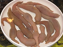
The species Urechis unicinctus is often used as bait by the fishing industry in Japan and Korea ; in Korea, China, Vietnam and in Chile, especially on the island of Chiloé , hedgehog worms are used as food.
swell
literature
- Wilfried Westheide , Reinhard Rieger: Special Zoology. Part 1: Protozoa and invertebrates. 2nd Edition. Spectrum Academic Publishing House, Heidelberg / Berlin 2007, ISBN 978-3-8274-1575-2 .
- Bernhard Grzimek (ed.): Grzimeks animal life . Bechtermünz Verlag, Augsburg 2000, ISBN 3-8289-1603-1 .
Individual evidence
- ^ R. Wehner , W. Gehring : Zoologie . 24th edition. Georg Thieme Verlag, Stuttgart, New York 2007, ISBN 978-3-13-367424-9 , pp. 199 .
- ↑ Estimates from: Wilfried Westheide, Reinhard Rieger (Ed.): Special Zoology Part 1: Protozoa and Invertebrates ; Gustav Fischer Verlag; Stuttgart, Jena & New York 1996, ISBN 3-437-20515-3 .
- ↑ From: Bernhard Grzimek (Ed.): Grzimeks Tierleben , Bechtermünz Verlag, Augsburg 2000, ISBN 3-8289-1603-1 .
