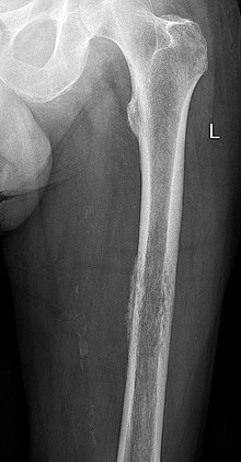
Lodwick classification
The Lodwick classification is used to assess whether a bone tumor is to be classified as benign (benign, e.g. juvenile bone cyst ) or malignant ( malignant , e.g. osteosarcoma ) on a normal X-ray (not on CT or MRI) . The classification takes place according to the severity of the osseous destruction pattern into classes I (benign) to III (malignant), with the subgroups Ia-Ic. Mixed forms are also possible.
The classification can also be used to assess the progression, especially to describe more aggressive behavior over the course of the disease. The classification is neither sensitive nor specific . Further imaging (such as scintigraphy , computed tomography or magnetic resonance tomography ) as part of a staging or biopsy can only be dispensed with if the findings are very clear .
The classification dates from the time before the introduction of computed tomography and magnetic resonance tomography and has lost its significance as a result of these procedures. In addition, it can only be used for solitary, focal bone changes, but not for generalized bone changes and only to a limited extent in the case of bone metastases . Nevertheless, it is well suited for describing the native radiological findings, since statements about the aggressiveness of the tumor are possible based on the X-ray morphology alone. On the other hand, it does not take into account the localization of the tumor in the bone, whether metaphyseal , diaphyseal or epiphyseal , whether cancellous , cortical or mixed, whether central or marginal.
The background to the classification is that in the case of slowly or non-growing bone tumors, such as a bone cyst , the tumor margin is usually clearly demarcated and a reactive surrounding reaction in the form of sclerotherapy, osteosclerosis or new bone formation takes place in order to avoid the mechanical loss caused by the less stable Cyst arises to compensate mechanically. In contrast, aggressive tumors, such as osteosarcoma , grow so quickly that they destroy the bone diffusely without a discernible edge and there is no time for reactive remodeling processes. Likewise , when the periosteum is lifted from the bone by tumor growth, it reacts with new bone formation, so that shell-shaped neoplasm strips ("onion skins") can be seen during rapid growth, while benign tumors do not penetrate to the periosteum or cause the periosteum to detach.
Destruction Pattern
- 1 - The tumor is clearly demarcated, often described as "map-like" or "geographic", and does not grow or grows slowly. It is usually found in the cancellous bone.
- 1A - The tumor remains limited to the cancellous bone with clear marginal sclerosis and sharp delimitation. The corticalis or periosteum are not penetrated. This pattern is typical in an enchondroma or osteoid osteoma . The non-ossifying bone fibroma , on the other hand, is a purely cortical defect.
- 1B - The somewhat faster growing active lesion has little to no sclerotic border, the contour is irregular with bulges. The cortex can also be affected, and a solid periosteal reaction is possible. Examples are the giant cell tumor , the predominantly epiphyseally localized chondroblastoma , the chondromyxoid fibroma , the osteoblastoma and also the juvenile and the aneurysmal bone cyst .
- 1C - This rapidly growing, partially infiltrative lesion shows mostly a "blurred" fuzzy contour, sclerosis is still possible. The cortex is usually eroded, and there may be a solid periosteal reaction. Examples are the low-grade chondrosarcoma and the aneurysmal bone cyst .
- 2 - This faster growing bone tumor can no longer be delimited geographically, but shows a "moth-eaten-like" destruction with a fuzzy border. There is usually no reactive sclerotherapy. The cortex is also destroyed, and the periosteum stands out like a bowl. This picture is typical of a high-grade malignant chondrosarcoma , a low-grade osteosarcoma and a fibrosarcoma .
- 3 - The tumor grows rapidly and destructively without respecting anatomical boundaries ("permeative"); reactive sclerotherapy is a big exception. The delimitation is usually no longer recognizable, and the periosteal reaction is "onion skin-like", partly interrupted, Codman triangles can form at the edges of the periosteal detachment . In adjacent soft tissue expansion soft tissue of the tumor can be prepared by the spicules be visible. Typical representatives are the highly malignant osteosarcomas and Ewing sarcomas .
literature
- F. Hefti: Children's orthopedics in practice . Springer-Verlag, Berlin 1997, ISBN 3-540-61480-X (p. 586)
- GS Lodwick, AJ Wilson: Determing growth rates of focal lesions of bone from radiographs. Radiology 1980; 134: 577-583
- Heuck, Wörtler, Vestring: Radiology of bone and joint diseases: primary and secondary bone tumors . Thieme Verlag, 1997, ISBN 3-13-107071-4
