Pemphigus vulgaris
| Classification according to ICD-10 | |
|---|---|
| L10.0 | Pemphigus vulgaris |
| ICD-10 online (WHO version 2019) | |
The pemphigus vulgaris (about ancient Greek πέμφιξ Pemphix "bubble, edema [on the skin]" and Latin vulgaris "usually"), even bubble addiction called, is a skin disease from the group of autoimmune blistering . It is characterized by blistering due to acantholysis of the lower layers of the epidermis . The reasons are IgG - autoantibodies against desmoglein 3 ( "pemphigus antigen", a protein component of the desmosome ) arranged in the intercellular spaces of the affected skin areas, as well as in the serum can be detected of the patients. This differs from pemphigus foliaceus , in which desmoglein-1 and the upper epidermal layers are affected.
Overall, the disease is rare and mainly affects people of middle and older age. A gender-specific accumulation is not known. The disease is more common in the Middle East than in Central Europe.
causes
It is an autoimmune disease . For unknown reasons, autoantibodies against desmoglein-3, a transmembrane cell adhesion molecule from the cadherin family, are formed. There are several hypotheses about the effects of autoantibodies:
- they disrupt the connection between the desmogle lines
- by triggering a signal, they cause apoptosis of the skin cells.
If there is a genetic disposition for autoimmune diseases, the disease can be triggered by various drugs , but also by viruses and UV radiation.
Symptoms
In over half of those affected, the disease begins with an attack on the oral mucosa . This creates rapidly bursting blisters that bleed easily and leave painful erosions. Subsequently, sagging blisters with a clear content appear in different places on previously healthy skin (spread to other mucous membranes, scalp and the entire integument , especially on places that are exposed to pressure and friction). The edge of the bubble expands eccentrically until the bubbles burst. Erosion occurs, which then becomes encrusted. The confluence of the blisters can affect large areas of skin where both crusts and intact blisters can be found next to each other.
Healing occurs from the center of the blisters. There are no scars here , but reactive hyperpigmentation can persist for a relatively long time on the affected areas. During acute phases it is possible to create a bubble on healthy skin by pushing sideways ( Nikolski phenomenon I positive ). Accordingly, the disease often manifests itself particularly strongly in the gluteal region , with the risk of secondary infections. In the case of extensive infestation, there is often a fever , loss of appetite and a general feeling of illness.
Diagnosis
The diagnosis of the disease is mainly based on the typical clinical picture and the positive Nikolski phenomenon (triggering of bubbles by pushing the slide or the possibility of moving existing bubbles by pushing sideways). Evidence is then provided by the presence of antibodies against desmoglein-3 in the serum.
Diagnostics include:
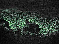
- Detection of autoantibodies bound in the skin in skin biopsies (direct immunofluorescence)
- Detection of autoantibodies circulating in the blood
- on biopsies of healthy skin or in the monkey esophagus in the spaces between the epithelial cells after incubation with patient serum (indirect immunofluorescence)
- in the ELISA with desmoglein-3 (also with desmoglein-1)
- Dermatohistopathology (suprabasal cleft or blistering)
- Tzanck test : detached, rounded keratinocytes are found on the base of the blister .

Differential diagnoses
- Pemphigus foliaceus
- Paraneoplastic pemphigus
- Bullous pemphigoid
- Dermatitis herpetiformis Duhring
- Hailey-Hailey disease
Histopathology
Acantholytic blisters are located suprabasally. The basal keratinocytes remain intact, and underneath the dermis is infiltrated by leukocytes . "Pemphigus cells" (rounded keratinocytes) are found in the lumen of the blisters.
therapy
The disease is mainly treated with the systemic administration of glucocorticoids . Initially, high doses have proven effective (1 to 3 mg prednisolone / kg body weight / day), which are gradually reduced until a minimum maintenance dose can be found. If this is above the so-called Cushing's threshold, immunosuppressants are also used to keep the long-term administration of glucocorticoids as low as possible (e.g. azathioprine, 2 to 2.5 mg / kg body weight / day, depending on the activity of the Thiopurine methyltransferase ).
According to recent studies, rituximab , a monoclonal antibody against the B-lymphocyte surface antigen CD20, e.g. T. in combination with immunoglobulin, effective.
Another therapeutic option is immune adsorption . This form of therapy is similar to plasmapheresis , whereby the separated blood plasma is passed through a protein A column. Most of the (IgG) antibodies are bound there. The plasma is then returned to the patient. Even if all other (necessary) antibodies are bound in addition to the autoantibodies that cause disease, the immune adsorption mainly has an effect on the autoantibodies for reasons that have not yet been clarified.
People with moderate to severe disease are treated as inpatients.
forecast
There are both acute and chronic courses. If left untreated, the disease is fatal after a few years. Since the use of glucocorticoids and immunosuppressants, the prognosis of the disease has improved significantly. Nowadays 10–20% of the causes of death are side effects as a consequence of long-term therapy with glucocorticoids and immunosuppressants, which means that 80–90% survive the disease due to the therapy. In the past, 100% lethality was due to illness due to fluid loss, superinfection and other complications of a destroyed skin barrier .
Web links
- Pemphigus Vulgaris Network
- Entry on pemphigus vulgaris in the Roche Lexicon online
- Dermis: Pictures of the pemphigus vulgaris
literature
- ↑ Preety Sharmaa, Xuming Maoa, Aimee S. Payne: Beyond steric hindrance: The role of adhesion signaling pathways in the pathogenesis of pemphigus . In: Journal of Dermatological Science , 2007, 48, pp. 1-14, PMID 17574391 .
- ↑ G. Rassner (Ed.): Dermatology: Textbook and Atlas . 7th edition. Urban & Fischer, 2002, ISBN 3-437-42761-X , p. 151 .
- ↑ a b c d Gernot Rassner: Dermatology: Textbook and Atlas . 8th, completely revised and updated edition. Elsevier, Urban & Fischer, Munich 2007, ISBN 978-3-437-42762-6 .
- ↑ A. Razzaque Ahmed, Zachary Spigelman, Lisa A. Cavacini, Marshall R. Posner: Treatment of Pemphigus Vulgaris with Rituximab and Intravenous Immune Globulin . In: New England Journal of Medicine . 2006; 355, pp. 1772-1779.
- ↑ I. Shimanovich, S. Herzog, E. Schmidt, A. Opitz, E. clinker, EB Bröcker, M. Goebeler, D. Zillikens : Improved protocol for treatment of pemphigus vulgaris with protein A immunoadsorption. In: Clinical and Experimental Dermatology . 2006; 31, pp. 768-774. doi : 10.1111 / j.1365-2230.2006.02220.x
- ↑ Daniel M. Peraza, Pemphigus vulgaris , MSD Manual, patient edition, April 2017; accessed on July 17, 2019.
