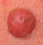
Pigment nevus
| Classification according to ICD-10 | |
|---|---|
| D22.9 | Pigment nevus |
| ICD-10 online (WHO version 2019) | |
A melanocytic nevus (sometimes also: Melanocytic or melanocytic nevus ) is limited, benign malformation of the skin , in contrast to other types of nevi from pigment-producing melanocytes is or related cell types and therefore generally has a brown or brownish color. There are numerous subspecies of pigment nevi (see → classification ).
Explanation of terms
Due to historical developments, there is some linguistic overlap with regard to the designation of pigmented nevi:
- In colloquial language , terms such as “birthmark” or “ mole ” are used to describe benign, brownish spots on the skin , but these cannot be applied exactly to dermatological terms. A “mole” usually corresponds to a lentigo or a nevus cell nevus , while the term “birthmark” is usually used as an umbrella term for all benign, circumscribed skin changes (technical term: “nevus”).
- In some sources pigmented nevi are also Melanozytennävi or melanocytic nevi called, which refers to the general descent from the melanocytic system. However, since not every pigment nevus is made up of normal melanocytes, these terms should not be used in order to avoid confusion with “melanocytic nevi” in the narrower sense.
- All types of pigmented nevi can also be referred to as pigment spots .
- Occasionally, the term “pigment nevus ” is used to describe a nevus pigmentosus (café-au-lait spot) or nevus spilus .
- In English usage, pigmented nevi are referred to as moles , hence the “new German” expression moles .
Cell types
Pigmented nevi can consist of the following cell types:
- In these nevi there are more normal melanin-forming dendritic melanocytes and produce more skin pigment ( melanin ). They are called melanocytic nevi and, according to their location in the skin layers, are further subdivided into epidermal melanocytic nevi and dermal melanocytic nevi .
- These cells are closely related to the melanocytes, but differ from them in the lack of dendrites, their spherical to spindle-shaped shape and their arrangement in nests. In addition, they can no longer release their pigment to the surrounding skin cells. The nevus cell nevi are also sometimes given the attribute "melanocyte", which then only refers to their melanocytic origin and does not mean that they consist of normal melanocytes.
- atypical cells
- Dysplastic nevi can arise from melanocytes as well as nevus cells that have lost their normal shape and have numerous cell atypias .
Classification
-
melanocytic nevi ("real" melanocytic nevi from normal melanocytes )
-
epidermal melanocytic nevi (located in the epidermis )
- Ephelids (freckles)
-
Lentigenes
- Lentigo simplex mole
- Lentigo solaris age spot
- Lentigo maligna can develop into malignant lentigo maligna melanoma.
- Nevus pigmentosus (café-au-lait-stain)
- Nevus spilus
- Becker nevus
-
dermal melanocytic nevi (located in the dermis )
- Mongolian spot
-
Naevus fuscocaeruleus
- Nevus Ota
- Ito nevus
- Naevus caeruleus (Blue nevus)
-
epidermal melanocytic nevi (located in the epidermis )
- Nevus cell nevi (nevi from nevus cells )
- Nevi from atypical melanocytes or nevus cells
However, since there is a subtype of malignant melanoma ( lentigo maligna melanoma) that has clear similarities and develops from lentigo maligna , medical / dermatological assessment of age spots is recommended.
Clinical significance
etiology
Nevi (pigmented or not) are generally based on developmental disorders in the embryonic stage and arise from postzygotic mutations in the genetic material.
Epidemiology
Nevus cell nevus and lentigo simplex (later in life also lentigo solaris ) are the most common nevi in humans; on average, a white-skinned adult has about 20 acquired nevus cell nevi (which, however, go through a cycle and can disappear again). Dysplastic nevi occur in approximately 5% of white-skinned adults.
clinic
Epidermal melanocytic nevi and junctional nevi are sharply circumscribed, brown spots at the level of the skin, while dermal melanocytic nevi are broad, raised, less dark structures that can protrude above the level of the skin. Also dysplastic nevi and melanocytic nevi may have raised portions, the latter depending on their development phase: Over time melanocytic nevi sink into the dermis , and thus lead to a more pronounced grandeur. On a nevus cell nevus there may be a hypertrichosis come.
For detailed descriptions of the subspecies of pigment nevi see there:
- Ephelids (freckles)
- Lentigenes (age spots, liver spots)
- Nevus pigmentosus (café-au-lait spots)
- Nevus spilus
- Becker nevus
- Mongolian spot
- Naevus fuscocaeruleus (nevus ota, nevus ito)
- Naevus caeruleus (Blue nevus)
- Nevus cell nevus
- Congenital nevus cell nevus
- Halonevus
- Spitz nevus
- Dysplastic nevus
forecast
The exact assignment of the different types of pigment nevi to the “birthmarks” of a person, and thus an assessment of the melanoma risk, can only be carried out by a dermatologist or another doctor experienced in dermatology.
Freckles (ephelids), café-au-lait spots (nevi pigmentosi) and the small-spotted lentigens ( lentigo simplex , lentigo solaris ) consist of normal melanocytes and do not pose a risk for the development of a melanoma . However, the risk of melanoma is directly related to the total number of nevus cell nevi . Dysplastic nevi can arise spontaneously from nevus cell nevi or occur more frequently as part of a "syndrome of dysplastic nevi (DNA)" and are the precursors of a true melanoma of the skin. Even congenital melanocytic nevi (large nevi that actually existed since birth) are potential melanoma precursors and should be treated.
literature
Guidelines
- S1 guidelines for melanocytic nevi of the German Dermatological Society (DDG) and the German Cancer Society (DKG). In: AWMF online (as of 2010)
Others
- Thomas B. Fitzpatrick, Klaus Wolff (ed.): Atlas and synopsis of clinical dermatology: common and threatening diseases . 3. Edition. McGraw-Hill, New York; Frankfurt a. M., 1998, ISBN 0-07-709988-5 .
- Ernst G. Jung, Ingrid Moll (Ed.): Dermatology . 5th edition. Thieme, Stuttgart, 2003, ISBN 3-13-126685-6


