
Skeletal scintigraphy
The bone scan , also Bone scintigraphy ( engl. Bone scan called), is a nuclear medicine imaging technique ( scintigraphy ), which serves for the detection of bone portions with an increased bone metabolism.
Areas with increased bone metabolism (“herds”) can be found, for example, in bone metastases in the context of cancer (e.g. prostate carcinoma , breast carcinoma ), but also in the healing zone of bone fractures or in inflammatory changes such as B. osteomyelitis or loosening of implanted endoprostheses (e.g. hip or knee joint prostheses).
description
Skeletal scintigraphy is a method with high sensitivity (approx. 94%) for the detection of bone damage ( osseous lesions ). Especially with bone metastases, which are usually characterized by increased (positive contrast), more rarely by reduced storage (negative contrast in pure osteolyses). The physiological principle is based on chemisorption . The 99m Tc -labeled bisphosphonates , such as oxidronic acid , accumulate on the bone surface. The degree of enrichment depends on various factors: regional blood flow, capillary permeability, osteoid content and bone surface, and in particular osteoblast activity. Technetium is bound to bisphosphonates with an affinity for bones such as hydroxymethylene bisphosphonate (HMDP), hydroxyethylene bisphosphonate (HEDP), methylenediphosphonate (MDP) or diphosphono-1,2-propanedicarboxylic acid (DPD). The usual activity in adults is in the range of 500 to 720 MBq .
If a circumscribed skeletal change is suspected, the examination is carried out as a multi-phase scintigraphy (e.g. three-phase scintigraphy). To this end, at the beginning of the "bolus" - injection made over the span of a minute a Sequenzszintigraphie with a recording per second (perfusion). Then the static early recordings (blood pool phase) of the affected skeletal area take place. After at least two hours, images ( scintigrams ) of the entire skeleton are made in ventral and dorsal projection (bone phase) and supplemented by target images of clinically or scintigraphically abnormal regions, possibly using the SPECT technique.
The principle of the investigation is that phosphate compounds are attached to the hydroxyapatite crystals of the bone depending on the blood flow and thus reflect the bone blood flow in the early investigation phases of scintigraphy and the bone metabolism in the subsequent later phases. If the blood supply is interrupted, a bone lesion does not change the bone metabolism or a bone is completely destroyed, no accumulation can be detected.
A radionuclide is injected intravenously for the examination . After a certain waiting time, the skeletal system can then be recorded with a gamma camera .
PET / CT using the tracer 18 F sodium fluoride is more efficient than skeletal scintigraphy with the aid of a gamma camera . Although the method is more complex and expensive, PET imaging is superior to scintigraphy in terms of resolution and sensitivity due to the fundamentally different technology of PET imaging . Outside of clinical studies or in non-high-risk patients, skeletal scintigraphy is used for routine diagnostics, for example when searching for bone metastases, as the method of second choice, especially when magnetic resonance imaging or PET methods are less readily available.
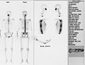
Full body picture of a patient ( lesion in the facial skull on the right).
Full body picture of a patient with Marie Bamberger Syndrome .

Full body picture of a 7 year old child. Normal findings; intensive enrichment in the growth plates .
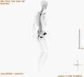
PET / CT in maximum intensity projection with 18 F sodium fluoride (NaF) in metastatic breast cancer. Bone metastases in the skull, spine, pelvis, ribs, left collarbone and right thigh.

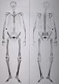
HDP skeletal scintigram of a patient with kidney cancer : The bone metastases in the cervical vertebrae 7 and in the lumbar spine (LWK 1 & 2) can only be guessed at due to the low resolution of the planar skeletal scintigraphy.

SPECT image of the HDP skeletal scintigram of the same patient; of the two metastases in the lumbar spine, only a single lesion can be seen; the lesion in the cervical spine is not shown.
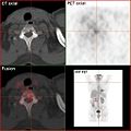
FDG- PET from the same patient: the osteolytic bone metastasis in the cervical vertebral body does not accumulate and can only be seen on CT. The two other bone metastases in the lumbar spine, on the other hand, are also shown in the FDG-PET.

Sodium fluoride PET-CT of the same patient: The osteolytic bone metastasis in the cervical spine shows an extreme uptake and is clearly recognizable. The two other bone metastases in the lumbar spine are also shown very well in the PET.


literature
- S1 guidelines for skeletal scintigraphy of the German Society for Nuclear Medicine (DGN). In: AWMF online (as of 2013)
Individual evidence
- ↑ J. Frey Schmidt, A. Stäbler: manual diagnostic radiology. Verlag Springer, 2005, p. 8, ISBN 978-3-540-26388-3 limited preview in the Google book search
- ^ JA Scott, EL Palmer: PET-CT of Bone Metastases. In: P. Shreve, DW Townsend (Ed.): Clinical PET-CT in Radiology - Integrated Imaging in Oncology. Springer Science + Business Media, LLC 2011 ISBN 978-0-387-48900-1 , Chapter 32
- ↑ FE Lecouvetemail et al .: Monitoring the response of bone metastases to treatment with Magnetic Resonance Imaging and nuclear medicine techniques : A review and position statement by the European Organization for Research and Treatment of Cancer imaging group. Eur. J. Cancer 50 (15) 2014, p. 2526.