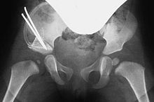
Acetabuloplasty
The term acetabuloplasty summarizes various surgical techniques that - under the generic term pelvic osteotomies - are used for the surgical treatment of hip dysplasia (HD) in childhood. These include some technically very similar operations, such as the osteotomy according to Lance, according to Pemberton or according to Dega. The Salter osteotomy also belongs - in the broader sense - to the group of acetabuloplasty, but the procedure is very different from all other procedures.
Basics
The pelvis , or rather the hip bone , is made up of three bones, the iliac bone , the pubic bone and the ischium . During growth , the connection points (growth plates) between the three bones remain open. They are only connective tissue , later flexibly connected to one another by cartilage tissue and only ossify at the end of the bony growth. The three growth plates meet in the later center of the acetabulum and form the Y-joint there.
In the case of hip dysplasia, the femoral head lacks the lateral (lateral) and front (ventral) roofing (also known as the pan orifice ). The "future" femoral head is not properly covered and threatened - depending on the severity of dysplasia - slipping up and dislocate (dislocate).
The principle of acetabuloplasty makes use of the still open Y-joint. The iliac bone is severed above the socket (osteotomy) so that the lateral socket can be swiveled down. The Y-joint is the pivot point. This principle is the basis of all acetabuloplasty techniques, only the procedure is different.
Aim of acetabuloplasty
The aim of this operation is to restore the lateral and ventral acetabular cavity in such a way that the femoral head is physiologically covered. The earlier the operation is performed (given the indication ), the greater the likelihood that the hip joint and femoral neck will grow normally.
The lateral roofing is measured at the so-called acetabular angle (AC angle) in the pelvic overview x-ray: An angle between a horizontal line through the Y-joints and a line along the cup cavity. In healthy newborns, the AC angle is around 25 °, around 15 ° at 6 years of age and 11–12 ° from the age of 12. The AC angle should also be corrected during acetabuloplasty according to these physiological values. One speaks of anatomical reconstruction .
Indications and contraindications
The main indications for acetabuloplasty is hip dysplasia. Surgical intervention is necessary if the HD can no longer be treated conservatively - i.e. with spreader pants / splint, spreader cast or reduction cast - or these treatment methods have failed. An absolute indication for acetabuloplasty is non-repositionable (non-repositioning) hip dislocation. If the indication for acetabuloplasty is made, the operation should be done as soon as possible.
The operation can be performed on babies from the first month of life if there are no other medical reasons against it. A joint-improving surgical procedure is recommended from an age of around one and a half years, since only then will the formation and strength of the bone allow an exact and clean implementation. In most cases, the previous conservative measures mean that the operation does not take place before the age of two at the earliest. Mild forms of hip dysplasia often show a favorable form, which is why in such cases an operative treatment plan is usually waived until the age of 3.
There are different opinions about the age up to which acetabuloplasty can be performed. It is crucial that the Y-joint must still be open, which makes the operation possible at an age of 12 or 13 years - i.e. until the Y-joint is closed. If the initial diagnosis is late (6 months of age and older), the indication for direct surgery is given, but conservative measures should nevertheless be considered, depending on the findings and severity.
Another indication is Perthes' disease (early childhood femoral head necrosis), whereby the Salter method is often used in conjunction with an intertrochanteric varization osteotomy (inward tilting / correction of the femoral neck with the aim of better centering the femoral head in the acetabulum). Acetabuloplasty is also used for rare neurological disorders (e.g. infantile cerebral palsy ) that lead to hip dysplasia or hip dislocation.
Contraindications are febrile infections , inflammatory processes in the area of the hip joint or pelvic bone and other general findings that have not yet been clarified. If the Y-joint is closed, the bony growth is complete and the femoral head is severely deformed, the operation can no longer be performed.
Diagnostics and diagnosis
The details of the clinical and imaging diagnosis of hip dysplasia or hip dislocation are explained in the article Hip Dysplasia.
Conventional x-ray images of the pelvis, so-called pelvic overview images and Rippstein images , are made preoperatively, both for the precise assessment of the joint misalignment and its severity, and for planning the operation itself . The Rippstein recordings are used for a more precise assessment of the femoral necks in a lateral projection.
anesthesia
The operation is performed under general anesthesia with intubation or a laryngeal mask . For pain therapy, the children are given a pain reliever - in the form of a suppository or as an injection - before the operation .
Operational sequence
In the case of dysplastic hip dislocation, a functional arthrography of the hip joint (s) can be performed first. Here the dislocation behavior and the degree of capsule overexpansion / damage can be precisely determined in the X-ray image. At this point at the latest, the decision can still be made as to whether or not an operation is necessary, and if so, which procedure is used.
Technique of acetabuloplasty
With all osteotomy techniques, only a small access is required between the inguinal fold and the iliac crest. The muscles are pushed apart here and the iliac bone is shown below. Shortly above the socket, the periosteum is detached and the iliac bone is exposed.
Pemberton osteotomy
At Pemberton, the iliac bone is notched about 5 mm above the socket under constant x-ray control with a flat chisel and the osteotomy is completed in the direction of the Y-joint. The socket fragment is now folded down and forward at the same time, also under X-ray control. In this way the most anatomical reconstruction of the socket is achieved.
Osteotomy according to Dega
Dega also carries out the osteotomy in the direction of the Y-joint, but uses special, curved chisels in order to achieve a detachment of the fragment that is as spherical as possible - adapted to the cup curve. Originally, Dega only lowered the socket fragment laterally, but today this is mostly modified with an additional ventral pivoting (forward), similar to the Pemberton method.
According to the gap created in this way, a suitable bone wedge (donor bone / bone bank see below) is sawn to size in both procedures and, under X-ray control, pushed into the gap. If necessary, the wedge can be fixed with an osteosynthesis wire (also called a Kirschner wire). For a trained surgeon, this operation takes about forty-five to sixty minutes.
Following the operation, a corresponding pelvic leg cast (modified Fettweiss cast) or an abduction orthosis is put on so that the femoral head is in the center of the socket during postoperative healing. The anesthesia is only ended afterwards.
The bone wedge
In children it is difficult to use their own bone in the sense of autologous (bone) transplantation . In the case of simultaneous intertrochanteric (between the protruding bones on the thigh bone), an attempt can be made to use the sloping correction wedge. In most cases, however, it is too small.
Today donor bones from in-house bone banks or from various manufacturers are mainly used. These are mostly femoral heads that are removed during endoprosthesis operations and donated to otherwise healthy patients who have given their consent. The donor bone are in certified thermal disinfection equipment disinfected and deproteinized. They are then in sterile containers - - at least minus 20 ° C. Cryopreserved . Alternatively, it is possible to sterilize donor bones in an autoclave under the influence of pressurized steam . Other sterilization methods , for example gamma-ray sterilization, are also used in industrial bone preparation.
All processing and disinfection / sterilization processes as well as the storage of the donor bones (deep freezing) are complex and are subject to strict requirements of the German Medical Association , the Medical Devices Act (MPG) and the Robert Koch Institute .
special cases
In rare, very severe cases, the femoral head can no longer be repositioned manually (overhead technique) (brought into the normal position). The femoral head has slipped so far and the joint capsule so overstretched and hypertrophied that the head cannot be repositioned back into the socket. Often, excess capsule material is deposited in the socket and additionally hampers reduction. In such cases, the femoral head must be repositioned in an open manner, i.e. by opening the joint itself. One speaks of "open hip adjustment". The capsule is opened in a circular manner, excess capsule and joint mucous membrane tissue is removed from the socket, the femoral head is repositioned and the capsule is gathered over the femoral head if necessary.
Complications
General complications
As with any surgery, vascular and nerve injuries can occur. The simple and safe access, as well as the short duration of the operation, keep blood loss to a minimum. Surgery on bones can lead to unexpected fractures that have to be treated directly during the operation.
Postoperatively, there can be further complications, such as wound infections and other wound healing disorders , arthritis of the hip joint or, in the worst case, sepsis .
Specific complications
Intraoperative injuries to organs or larger blood vessels are not described in the literature. Injuries or irritations of the lateral femoral cutaneous nerve (sensitive inguinal and femoral nerve ) can occur, but are mostly reversible.
A postoperative failure or collapse ( sintering ) of the bone wedge can make a new operation necessary. It can also happen that the bone wedge has not been anchored firmly enough and it loosens postoperatively from the osteotomy gap, which usually leads to the dissolution ( lysis ) of the wedge. The pelvic leg plaster can cause pressure damage or nerve irritation.
Follow-up treatment and rehabilitation
The immobilizing cast (retention cast) that is put on after the operation is usually left for six weeks. A control X-ray can be taken after one and after another two weeks. After the six weeks, a cast should be changed, which usually requires another anesthetic. The plaster of paris is worn for a total of three months and then replaced by a splint treatment. Continuous follow-up examinations and controls are imperative. The older the child, the longer the follow-up treatment will take.
Success rates
There are a few studies that describe the results and success rates of acetabuloplasty with clinical and radiological follow-up examinations. One study even describes the results in the medium-term range. She examines eighty-three children (a total of 125 operated joints) ten years after acetabuloplasty using the Pemberton technique. Not only the results of the cup correction, but also the success of the use of sterilized foreign bone are examined. The follow-up examinations relate to the - already mentioned - AC angle, the osseous structure of the foreign bone wedge, the gait and movement development and the postoperative complaint situation. The overall result of this follow-up examination is extremely positive: Over 96 percent of the patients (and their parents) examined afterwards assess the result of the operation as good or very good. Only one patient rated the procedure as bad.
The earlier HD is detected, the easier and more effective the therapy. Since hip sonography is now standard in newborns ( U3 ), most dysplasias can be detected very early and treated accordingly.
history
The history of surgical therapy for cup misalignments begins at the end of the 19th century. In 1891, F. König tried for the first time in Berlin to fold down a periosteal bone scale in the lateral acetabular cavity. Albee (1915) and Jones (1920) used this concept and developed from it the basic form of today's acetabuloplasty. They deposited bone chips from the shin into the osteotomy gap. In 1924, Spitzy tried a method that was unsustainable. He fixed bone chips (also from the shin) to the dysplastic edge of the socket. They should form a new pan bay as they grow. More than 50 years later this method was taken up again. The resulting operation is still used today as a "shelf plastic". In 1925, the pediatric surgeon P. M. Lance in France took up the modifications by Albee and Jones again: He fixed a bone wedge in the osteotomy gap. To date, this technology has been modified, further developed and improved many times. Pemberton and Dega aren't the last in this line. Various surgeons and clinics are still developing this technique today. For example, they try out the use of artificial bones and minimally invasive techniques for access.
Literature and Sources
- AB Imhoff, R. Baumgartner: Checklist Orthopedics . Thieme 2006. ISBN 3131422815
- Breusch, Mau, Sabo: Clinical Guide Orthopedics . Elsevier 2006. ISBN 9783437224713
- Klaus Buckup , LC Linke, W. Cordier: Pediatric Orthopedics . Thieme 2001. ISBN 3136976029
- J. Duparc: Surgical Techniques in Orthopedics and Traumatology . Bd. Pelvic ring and hip. Elsevier 2005. ISBN 3437225561
- F. Hefti, R. Brunner: Pediatric orthopedics in practice . Springer 2006. ISBN 978-3540614807
- R.-P. Meyer, A. Gächter: Hip surgery in practice . Springer 2005. ISBN 978-3540227182
- Results of acetabuloplasty using the modified Dortmund technique (dissertation by Robert Bonmann 2003) (PDF file; 1.38 MB)
- CJ Wirth: Orthopedics and orthopedic surgery . Bd. Pelvis / hip. Thieme 2004. ISBN 9783131262219
Individual evidence
- ↑ a b c d e Dissertation on the results of acetabuloplasty (PDF; 1.5 MB)
- ↑ a b C.J. Wirth: Orthopedics and orthopedic surgery . Bd. Pelvis / hip. Thieme 2004.
- ↑ a b c d e A. B. Imhoff, R. Baumgartner: Checklist Orthopedics . Thieme 2006. ISBN 3131422815
- ↑ a b c d e f g R.P. Meyer, A. Gächter, U. Kappeler: Hip surgery in practice . Springer 2005. ISBN 9783540227182
- ↑ a b c K. Buckup, LC Linke, W. Cordier: Kinderorthopädie . Thieme 2001. ISBN 3136976029
- ↑ a b James G. Jarvis: Dega-Osteotomy in Hip Dysplasia . In: Operative Orthopedics and Traumatology. Volume 10, Number 2 / June 1998. pp. 117-124. ISSN 0934-6694 .
- ↑ P. Pemberton: Pericapsular osteotomy of the ilium for treatment of congenital subluxation and dislocation of the hip . J Bone Joint Surg 47-A, 65-86. 1965.
- ↑ a b c F. Hefti, R. Brunner: Pediatric orthopedics in practice . Chapter Hip Dysplasia / Acetabuloplasty. Springer 2006
- ↑ German Medical Association: Guidelines for managing a bone bank (PDF) ( Memento from December 16, 2015 in the Internet Archive )
- ↑ Steffen Schröter: Biomechanical properties of human cancellous bone after gamma sterilization versus heat disinfection with the Lobator . Dissertation at the Medical Faculty of the Eberhard Karls University in Tübingen. 2005.
- ^ FH Albee: The bone graft wedge. NY Med 52, 433-441 (Am J Med Sci 149, 313-325).
- ^ E. Jones: The operative treatment of irreducible paralytic dislocation of the hip. Amer J Orthop Surg 18 (1920).
- ^ H. Spitzy: Artificial pan roof formation . 1924. Z Ortop 43, 284-294.
- ^ PM Lance: Constitution d'une butee osteoplastique dans les luxation et subluxation congenitales de la hanche . 1925. Press Med 33, 945-948.
- ↑ P. Pemberton: Pericapsular osteotomy of the ilium for treatment of congenital subluxation and dislocation of the hip. 1965. In: J Bone Joint Surg Am 47-A, 65-86.
- ↑ W. Dega: Development and clinical significance of the dysplastic acetabulum . 1973. Orthop 2, 202-218.


