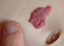
Hemangioma
| Classification according to ICD-10 | |
|---|---|
| D18.0 | Hemangioma, any location |
| ICD-10 online (WHO version 2019) | |
A hemangioma , also haemangioma or strawberry patch called, is one of embryonic tumor with endothelium - proliferation and secondary education of the vessel lumen . Typically, hemangiomas are still very small at birth and then increase significantly in size in some cases (approx. 10 percent), especially in the first year of life. Some forms appear after the 3rd decade of life.
Frequency and location
The incidence of hemangiomas is 3–5 percent in infants, premature babies are affected up to 10 times more often, thus the hemangiomas represent the most common tumors in childhood. Hemangiomas are two to three times more likely to occur in girls than in boys.
Hemangiomas can occur all over the body and also in internal organs, but in 60 percent of cases they occur in the head and neck area (formerly known as head blood lumps ). About a third of the hemangiomas are located in the liver. From angiomatosis is called the infestation of large areas or entire limbs.
Hemangiomas are usually congenital, show different growth tendencies and sometimes regress on their own. As a rule, they do not degenerate.
origin
The origin of the infantile hemangiomas is unknown. However, there is evidence that the vascular wall cells of the capillary vessels have a certain similarity with the placenta in their expression of genes. The self-limiting growth of the hemangiomas could therefore reflect the limited growth time of a placenta.
Forms, course and therapy
Capillary hemangioma
The capillary hemangioma (lat. Haemangioma capillare ) consists of capillary blood vessels and accounts for 30 to 40% of all vascular tumors. It can show up on the skin as a bright red raised vascular abnormality. Capillary hemangioma is quite common with one in 200 births and usually occurs shortly after birth. It usually grows in the first few months of life. More than 70 percent of capillary hemangiomas disappear almost completely with scarring by the age of 7. No therapy is required for uncomplicated hemangiomas. If the hemangioma is on the face or in the ano-genital area, early therapy should be given, otherwise if there is a clear tendency to growth. It cannot be predicted whether a capillary hemangioma will degenerate malignantly, but the speed is then high and can appear over a large area. This can lead to blindness in the eye area, deformities in the nose area or permanent changes in the lip area. The therapy is carried out with laser therapy, with cryogenics (cold therapy), in the case of very large capillary hemangiomas, treatment is carried out with steroids , chemotherapeutic agents and, since 2008, with β-blockers ( propranolol ). Surgical intervention is reserved for individual cases. Rare association with Kasabach-Merritt syndrome .
histology
- lobular vascular pattern of the vascular, canalized tumor
- focal microthrombi
- medium-caliber arborizing vessels
Cavernous hemangioma
→ Main article: Cavernoma
The cavernous hemangioma (lat. Haemangioma cavernosum ) or cavernoma is a light red to purple vascular malformation with large, cavernous vascular cavities. Sometimes it is already present at birth, but more often occurs in the first few days of life. Cavernous hemangiomas can be subdivided into cutaneous, cutaneous-subcutaneous and subcutaneous hemangiomas. The rate of regression of cavernous hemangiomas is around 80 percent.
Cavernous hemangiomas can contain arteriovenous vascular malformations and therefore bleed profusely. Further complications can be superinfection , necrosis and consumption coagulopathy (see Kasabach-Merritt syndrome ). Large cavernous hemangiomas can affect limb growth in children and should then be addressed therapeutically. Therapeutically, u. a. cryotherapy for small, flat hemangiomas. In the case of larger hemangiomas, the focus is on laser therapy . Cavernous hemangiomas are nowadays usually treated therapeutically because they are easy to treat and have a clear growth tendency. Cavernous hemangiomas in the liver are usually directly subcapsular, are usually smaller than 2 cm and are bluish in color. They can also occur in the brain.
Grape or berry-shaped hemangioma
Haemangioma racemosum is the so-called tendril angioma and occurs primarily in the head, neck and back area. It consists of tortuous and enlarged artery or vein and probably represents a true neoplasm ( neoplasia represents). The congenital hemangioma racemosum the retina occurs in so-called Bonnet-Dechaume-Blanc syndrome on.
Sclerosing hemangioma
This hemangioma ( Haemangioma cirsoideum ) occurs mainly in middle adult life. It appears as a relatively mobile, slowly growing lump up to 1 cm in size in the dermis and subcutis . The tumor is, as it were, a well-vascularized dermatofibroma and histologically, hemosiderin- positive deposits can be found. Therapy is excision .
Haemangioma planotuberosum
The haemangioma planotuberosum is a flat, raised, bulging, elastic, blue-red vascular tumor that originates from the vessels below the papillary body of the skin. Histologically, the tumor consists of capillary sprouts and immature endothelia.
Hemangioma of the eye socket
The hemangioma of the eye socket (lat. Orbita , therefore also: orbital hemangioma) in contrast to the lid hemangioma. The orbital hemangioma is the most common benign tumor in adults in the orbit, an area surrounded by skull bones with comparatively few tumors. Relatively often there is a corresponding incidental finding in skull examinations. The first symptom is a change in the position of an eyeball . Otherwise, angio magnetic resonance imaging and computed tomography of the paranasal sinuses are required for diagnosis .
A special feature of the orbital hemangioma is its occurrence in middle age. No gender preference can be determined; In one collective, around 65 percent of the findings were made by women. The tumor usually leads to slowly progressing exophthalmos (74 percent), reduced visual acuity (51 percent) and choroidal folds (32 percent), hyperopia, and diplopia . Therapy is only used for increasingly progressive tumors that are causing symptoms. The form of therapy here is surgical extirpation after previous obliteration . The differential diagnosis (DD) must be observed! Smaller and asymptomatic hemangiomas with typical clinical and radiological findings should be left in place because of the lack of benefit and possible surgical complications. Periodic checks are then sufficient. There are also recommendations for early surgical intervention in the absence of symptoms in order to obtain a definitive histopathological diagnosis and to prevent potential damage from further growth.
Hemangiomas of the spine
A vertebral hemangioma is the most common benign tumor of the spine and affects up to 10% of the general population. It is mostly discovered accidentally when examining the thoracic spine using imaging procedures such as CT or MRI and is particularly often found between the T3 to T9 vertebrae. Usually symptom-free, it generally does not require treatment. The most common shape and size roughly correspond to the one shown, but can also include the entire vertebral body.
Hemangiomas of the liver
→ Main article: Liver hemangioma
Hemangiomas are the most common (benign) neoplasms of the liver. They are often discovered as a chance diagnosis in sonography . They are not dangerous. Hemangiomas located only on the surface of the liver can rupture and bleed. There is no degeneration. However, deeper hemangiomas can lead to an intrahepatic obstruction of the drainage of the bile.
Syndromes
Kasabach-Merritt syndrome
→ Main article: Kasabach-Merritt syndrome
This syndrome leads to the development of benign, cavernous and large to giant hemangiomas. Mostly it affects women. Thrombocytopenia can develop due to localized disseminated intravascular coagulation (DIC) with thrombus formation within the hemangioma . Kasabach-Merritt syndrome does not occur in classic infantile hemangiomas, but rather in tufted angiomas and Kaposi-like hemangioendotheliomas.
See also: phacomatoses
Differential diagnosis
In the case of vascular malformations (vascular anomalies), a distinction must be made between hemangiomas and vascular malformations (vascular malformations) . Hemangiomas are often only discreetly present immediately after birth and can then increase in size significantly in the first few months of life. In contrast, vascular malformations often already exist at birth and become larger in proportion to body growth.
This distinction is important insofar as the treatment of hemangiomas must be carried out immediately if there is evidence of growth. In the case of vascular malformations, conservative therapy is also an option, depending on the case .
A differential diagnosis can also represent the rare Cobb syndrome .
etymology
The word is derived from the ancient Greek αἷμα haima "blood" and ἀγγεῖον aggeion [pronounced angeíon with an emphasis on ei ] "vessel". With the suffix -om (also -oma , -ma ) ends of many in Ancient Greek Neutra ; In medicine, in particular, technical terms are used to denote tumors (see tumor nomenclature ).
literature
- H. Ric Harnsberger, Patricia A. Hudgins et al. a .: head and neck. The Top 100 Diagnoses ; PocketRadiologist; Jena: Elsevier, Urban & Fischer, 2004; ISBN 3-437-23600-8 ; Philadelphia: Saunders, 2004; ISBN 0-7216-9697-X .
- Therein: H. Christian Davidson and Richard H. Wiggins, pp. 123-137.
- Wolfram Wermke: Liver diseases: textbook and systematic atlas ; Cologne: Deutscher Ärzte-Verlag, 2006; ISBN 978-3-7691-0433-2 .
- Ulrike Ursula Ernemann, J. Hoffmann, E. Grönewäller, H. Breuninger, H. Rebmann, C. Adam, S. Reinert: Hemangiomas and vascular malformations in the head and neck area. Review article of the interdisciplinary consultation hour for vascular anomalies at the University Hospital Tübingen ; in: Radiologe 43 (2003), pp. 958-966.
- V. Petrovici: hemangiomas and vascular malformations ; in Serge Krupp (ed.): Plastic surgery - clinic and practice; Landsberg / Lech: ecomed, 2000; ISBN 3-609-76210-1 .
- JB Mulliken, AE Young: Vascular Birthmarks: Hemangiomas and Malformations ; Philadelphia: Saunders, 1988.
- S2k guideline hemangiomas in infancy and early childhood of the German Society for Pediatric Surgery (DGKCH). In: AWMF online (as of 2012)
- V. McAllister, B. Kendall, J. Bull: Symptomatic vertebral haemangiomas ; Brain: a Journal of Neurology, 1975, pp. 71-80
Web links
- Photos of hemangiomas on DermIS
- CM Barnés, et al .: Evidence by molecular profiling for a placental origin of infantile hemangioma. In: Proceedings of the National Academy of Sciences . Volume 102, Number 52, December 2005, pp. 19097-19102, ISSN 0027-8424 . doi : 10.1073 / pnas.0509579102 . PMID 16365311 . PMC 1323205 (free full text). (English)
- Liver sonogram
- Macroscopic specimen
- Histology of a hemangioma
- Vertebral hemangioma
Individual evidence
- ↑ M. Sand, D. Sand, C. Thrandorf, V. Paech, P. Altmeyer, FG Bechara: Cutaneous lesions of the nose. In: Head & face medicine Volume 6, 2010, p. 7, ISSN 1746-160X . doi : 10.1186 / 1746-160X-6-7 . PMID 20525327 . PMC 290354 (free full text). (Review).
- ↑ H. Hamm, PH Höger: Skin tumors in childhood. In: Dt. Ärzteblatt 108, 20, 2011.
- ↑ Dr. Alice Martin: Skin Lexicon: Hemangioma (blood sponges) - symptoms. Dermanostic, accessed on May 19, 2020 (German).
- ↑ Léauté-Labrèze et al .: Propranolol for severe hemangiomas of infancy. N Engl J Med. 2008; 358 (24): 2649-51. PMID 18550886 .
- ↑ McNab, 1989.
- ↑ JW Henderson, GM Farrow, JA Garrity, et al. a. (1990) and A.-J Lemke, I. Kazi, et al. a. (2004).
- ^ Wilhelm Gemoll : Greek-German school and hand dictionary. Munich / Vienna 1965.


