Thigh bone
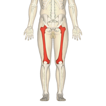
The thigh bone , in medical jargon Os femoris or the femur for short , is the strongest long bone and forms the bony basis of the thigh . It is the longest bone in the human body .
anatomy
head
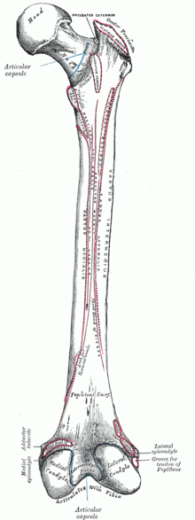
At the upper end of the thigh bone is its head ( caput ossis femoris ), which forms a connection with the pelvic bones and thus the hip joint with an approximately spherical joint surface . The head has a slight depression on its central circumference, the so-called femoral head pit ( Fovea capitis femoris ). It is the passage point for a ligament ( ligamentum capitis ossis femoris ), which surrounds the artery that supplies the femoral head.
neck
On the thigh bone further down (distal), the neck ( collum ossis femoris ) connects to the head . There are two protrusions on it:
- laterally the large roll mound ( greater trochanter ). It serves as an attachment to the gluteal muscles ( gluteus medius and gluteus minimus , piriformis , obturatores and gemelli muscles ).
- in the middle is the small hillock ( trochanter lesser ). It serves as the insertion of the iliacus muscle and the psoas major muscle .
The two rolling hills are connected on the abdomen (ventral) by a flat, rough line ( Linea intertrochanterica ) and on the back (dorsal) by a sharp ridge ( Crista intertrochanterica ). There is a depression ( fossa trochanterica ) between the large roll mound and the thigh bone . It serves as a base for several muscles ( internal obturator muscle , superior gemellus muscle , inferior gemellus muscle and external obturator muscle ).
Some mammals (e.g. horses and rabbits ) also have a third rolling mound ( trochanter tertius ).
Misalignments of the femoral neck are:
- Coxa vara - reduced CCD angle
- Coxa valga - enlarged CCD angle
- Coxa antetorta - larger than normal anteversion
- coxa retrotorta - Antetorsion of the femoral neck less than 0 °
shaft
The femoral shaft ( corpus ossis femoris ) begins below the rolling hills . It is almost cylindrical, but noticeably curved forward. In humans, a rough longitudinal line ( Linea aspera ), which consists of two ridges ( labium mediale and labium laterale ), runs across the posterior surface . - In animals, these “lips” are delimited by a rough surface ( facies aspera ). - In the middle of the shaft of the thigh bone, the two ridges lie close together, at both ends they diverge from each other (diverge). The lateral ridge widens towards the top (proximal) into an elongated roughness ( gluteal tuberosity ). The central bar runs flat into the line between the rolling hills and ends directly under the small rolling hill, which is directly reached by a second, parallel line ( Linea pectinea ). Further below, the two ridges diverge into two further lines ( Linea supracondylaris medialis and Linea supracondylaris lateralis ), which delimit an almost flat, triangular bone area ( Facies poplitea ). The lines and ridges serve as starting points for the pre-accession muscles ( adductors ).
Lower end
The thickened lower end of the thigh bone bears two strongly outwardly curved ( convex ) articular knots ( condylus medialis and condylus lateralis ). Together with the shin plateau, they form the knee joint . The two knots are separated from each other by a pit ( intercondylar fossa ). This is bounded at the back by a flat bone line ( Linea intercondylaris ). The cruciate ligament cavity (also known as the intercondylar notch ) is located between the two knots . At the front, the articular knots unite to form a common, transversely inwardly curved ( concave ), sagittal outwardly curved joint surface ( facies patellaris ) for connection with the kneecap ( patella ). The knots each have an attachment that protrudes only slightly ( epicondyle medialis and epicondyle lateralis ). The lateral attachment has a furrow ( sulcus popliteus ) on its side .
angle
CCD angle
The angle between the thigh neck (collum) and the bone shaft is called the center-collum-diaphyseal angle ( CCD angle for short ).
The CCD angle decreases from 137 ° in the 4th to 5th embryonic month to 129 ° in the 9th month of pregnancy and reaches 137 ° again around birth. The angle for infants is 145 °, for children about 140 °, from puberty onwards 130 ° for adults about 126 ° and for old people about 120 °.
Since the hip joint has to transfer large forces, the position of the joint socket, for example, must also be included in an assessment of the axial relationships. Physiologically, people with steep joint sockets also have larger CCD angles. With pathologically high values for the CCD angle, the clinical picture of the knock knees ( coxa valga ) is available (for example, in the case of immobility after muscle paralysis), since the body load does not exert any mechanical pressure on the femoral neck. A bow-leg ( coxa vara ) occurs when the thighbone neck is subjected to too much stress . This can be the case with a reduced resistance and thus an increased resilience of the bone (for example in rickets ).
Torsion angle
The torsion angle (syn. Anteversion angle or anteversion angle ) is understood as the rotation of the transverse axes of the distal and proximal femur end. The distal end of the femur is rotated inwards by approximately 12–20 degrees (in the direction of the median plane ) compared to the proximal end . This angle is larger in the child's bone than in the adult (see: Naiadensitz ).
Diseases
Fractures of the thighbone are relatively common, with the femur neck ( femoral neck fracture ) and shaft particularly affected.
The joint surface of the femoral head can be damaged by signs of wear and tear such as osteoarthritis , mechanical stress, injuries or inflammation . It can be replaced by a metal prosthesis (possibly together with the femoral neck) through an operation .
The articular surfaces of the two knots ( lateral condyle and medial condyle ) on the side of the knee joint are also often damaged, so that treatment (e.g. surgical replacement) may be necessary.
Individual evidence
- ^ Peter Matzen: Pediatric Orthopedics. Urban & Fischer Verlag, Munich / Jena 2007.
- ↑ What is the femoral head? Anatomical details-femoral head | elsetech. Retrieved March 12, 2020 .
- ^ Rolf Bertolini : Systematic anatomy of humans. Volk und Gesundheit, Berlin 1979 and Fischer, Stuttgart 1979, ISBN 3-437-00375-5 , pp. 110-125.


