AV block
| Classification according to ICD-10 | |
|---|---|
| I44.3 | Other and unspecified atrioventricular block |
| I44.0 | 1st degree atrioventricular block |
| I44.1 | 2nd degree atrioventricular block |
| I44.2 | 3rd degree atrioventricular block |
| ICD-10 online (WHO version 2019) | |
An AV block (short for atrioventricular block , also known as atrioventricular conduction disturbance and disorder of the atrioventricular excitation propagation ) is a common heart rhythm disorder . The conduction of excitation between the atria and the ventricles at the atrioventricular node (AV node) of the heart is delayed, temporarily or permanently interrupted.
Lighter forms of AV block can go unnoticed and do not require treatment. More severe forms lead to a heartbeat that is too slow ( bradycardia , bradyarrhythmia ). In extreme cases, the chambers can even come to a complete standstill, which then requires emergency medication and pacemaker treatment .
to form
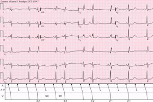
All forms of the AV block can be detected using an electrocardiogram (EKG).
1st degree AV block
The conduction of excitation is delayed and the ventricles of the heart begin to contract too late. This disturbance can be recognized by a lengthening of the PQ time (over 200 ms) in the EKG or by a shortening of the distance between the E and A waves in the echo that arise when the left ventricle is filled . Subjectively, it goes unnoticed and usually does not require any treatment.
Second degree AV block
The excitation line and thus the chamber action is partially canceled. There are several options:
- The PQ time is getting longer. Eventually it becomes so long that an atrial excitation is no longer transmitted and a single ventricular contraction fails. The subsequent atrial excitation is transmitted normally again. Then the PQ time extension begins again ( Wenckebach period ). The scientific definition of a Wenckebach block states that a P-wave is not transferred and the PQ-time of the P-wave before the failure is longer than the PQ-time of the P-wave after the failure.
- Sudden and unexpected failure of a ventricular action on atrial excitation without the PQ interval having to have been extended beforehand. Only every 2nd, 3rd or 4th atrial action can regularly be transferred to the ventricle (2: 1 or 3: 1 or 4: 1 block). This block form, which is mostly located in the bundle of His, is known as the Mobitz block . The prognosis is less favorable compared to the Wenckebach block, as there is a risk that the rhythm will transition into a total AV block.
In the English-speaking world, the Wenckebach block is also referred to as Mobitz Type I , the Mobitz block as Mobitz Type II or Hay .
AV block III. Degree
Complete failure of the conduction between the atrium and ventricle ( AV dissociation ). The ventricle stops or beats in a slow (bradycardic) substitute rhythm asynchronously to the atria, the AV node known as the secondary pacemaker (with approx. 40–50 pulses per minute) or the tertiary pacemaker (bundle of His, Tawaraschenkel and Purkinje fibers with a frequency of about 20-30 pulses per minute). An AV block III. Grades can with normal QRS complexes ( AV escape rhythm ) or widened QRS complexes ( ventricular escape rhythm ) occur.
A total AV block (complete atrioventricular dissociation) is a typical indication for implantation of a pacemaker .
Causes and frequency
Fröhlig names the following causes:
Congenital AV block
Congenital AV block is rare , with an incidence of 1 in 20,000. The causes are about one third each
- congenital heart defects ,
- idiopathic , possibly missing fusion of the conduction system or
- inflammatory, for example in the case of an autoimmune disease in the mother.
Acquired AV block
A variety of causes can lead to AV obstruction. The majority of cases are due to chronic degenerative changes (possibly) in the context of cardiac diseases. 40 to 60% of the cases are caused by idiopathic fibrosis (possibly). The most common reversible cause is medication . All antiarrhythmic drugs that delay conduction can cause AV blockages, especially if overdosed or in combination. Cardiac glycosides and beta blockers are the most common triggers in everyday clinical practice . Damage caused by medical interventions or myocardial infarction (see below) can be reversible.
Chronic degenerative
- Heart muscle disease
-
Infections
- Endocarditis
- Lyme disease
- Chagas disease
- other bacterial , viral , rickettial , fungal infections
- Neuromuscular Diseases
|
Excitation conduction system (schematic, in humans) 1 sinus node - 2 AV nodes Important structures are |
- infiltrative diseases
- Neoplastic disease
- Collagenoses
- Rheumatoid arthritis
Idiopathic
- Lev disease : fibrosis of the proximal conduction system with bilateral bundle branch block
- Lenègre's disease (or syndrome): Fibrosis that progresses from distal through the middle parts of the conduction system to proximal to total AV block, which is more likely to occur in younger people.
Other injury
- Circulatory disorder of the AV node
-
Cardiac surgery
- Heart valve replacement
- Ventricular septal defect occlusion
- Septal myectomy for HOCM
- Coronary artery bypass
-
Radio frequency ablation
- AV node, for example, in therapy-resistant tachycardic atrial fibrillation
- Accessory pathway in AV node reentry tachycardia
- Endocardial radiofrequency ablation of septal hypertrophy in HOCM
therapy
- Medication with atropine or orciprenaline only briefly to bridge the gap until the definitive supply with a pacemaker . Long-term therapy with medication is unsafe, has many side effects and is considered out of date.
CAVE: In the second-degree Wenckebach AV block, atropine may be used if there is severe bradycardia. In the Mobitz type, atropine should not be given, however, as it deteriorates with the risk of total AV block.
- Passage or permanent pacemaker in symptomatic patients of all types of block and in asymptomatic patients with AV block III. Degree or II degree type Mobitz.
See also
Individual evidence
- ^ Herbert Reindell , Helmut Klepzig: Diseases of the heart and the vessels. In: Ludwig Heilmeyer (ed.): Textbook of internal medicine. Springer-Verlag, Berlin / Göttingen / Heidelberg 1955; 2nd edition, ibid. 1961, pp. 450-598, here: pp. 571-574 ( conduction disorders ).
- ↑ Joseph Wartak: EKG practice. 2nd Edition. Thieme, Stuttgart 1982, ISBN 3-13-540502-8 , p. 124.
- ↑ G. Fröhlig et al. (Ed.): Pacemaker and defibrillator therapy . 1st edition. Thieme, Stuttgart / New York 2006, ISBN 3-13-117181-2 .
- ↑ Guidelines of the German Society for Cardiology, 2005: Pacemaker Therapy


