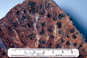
Emphysema
| Classification according to ICD-10 | |
|---|---|
| J43 | Emphysema |
| J44 | Other chronic obstructive pulmonary disease - with emphysema |
| ICD-10 online (WHO version 2019) | |
As pulmonary emphysema , emphysema (the lungs) or (chronic) pulmonary emphysema , an irreversible hyperinflation of the smallest air-filled structures ( alveoli , alveoli ) of the lung , respectively. It is the common endpoint of a number of chronic lung diseases .
Pathogenesis

One theory is that various pollutants (tobacco smoke, silicates, fine and quartz dust) or the body's own proteases (in the case of alpha-1 antitrypsin deficiency ) cause inflammatory changes in the lung tissue. The following collection of leukocytes releases proteases, including elastases . These destroy the elastic fibers of the lungs. Furthermore, oxygen radicals inactivate alpha-1-antitrypsin , which normally inactivates elastase and other proteases. In addition, the external pressure on the bronchioles (small bronchi) increases, which then collapse when you exhale , so that the air contained in the alveoli remains trapped ( trapped air ). The result is an overinflation of the lungs with an increased air content in the lungs. But there are also other guesses.
This means that less used air can be exhaled and less fresh air can flow in. In extreme cases, previously functional alveoli become large, non-functional "emphysema bladders". Endogenously persistent bronchial asthma can also lead to pulmonary emphysema.
Ultimately, the lungs lose their elasticity and the air they contain cannot escape completely.
Symptoms
Generally lies with emphysema due to the reduced gas exchange area, a chronic respiratory distress before, at first only during exercise, at a later stage even at rest ( dyspnea at rest ). The reduced oxygen content of the blood may be indicated by bluish-red discoloration ( cyanosis ) of the lips, fingertips and toe tips.
The circumference of the chest increases (barrel chest with horizontal ribs). In the late stage, there is a (right) heart strain .
In the X-ray image ( chest X-ray ) pulmonary emphysema shows up in a reduced density of the lung tissue . In addition, a teardrop heart can be formed.
Classification
Anatomically, the air paths branch off and on, until finally in the so-called alveoli end (alveoli) in which then finally the gas exchange takes place (figuratively largely like a grape present). The alveoli are attached to a small bronchus (several bronchioli respiratorii converge to form a bronchiolus terminalis). The pulmonary emphysema largely takes place in this area, i.e. the endings of the airways.
Pulmonary emphysema (formerly also called pneumonia ) can be classified as follows:
- Congenital lobar emphysema , rare congenital misalignment with massive overinflation of the lung lobes through a valve mechanism due to misalignment of the bronchi or the cartilage braces, lumen-relocating masses or an unknown cause.
- Primarily atrophic emphysema (normal age emphysema or senile emphysema ).
-
Secondary emphysema , d. H. as a result of an underlying disorder:
- Centrilobular (centroacinares) emphysema : It typically arises from chronic obstructive pulmonary disease (COPD). This type is found primarily in the upper lobes of the lungs . Initially the bronchioli respiratorii (the fine ramifications of the bronchial tubes that lead from the bronchioli terminales practically directly to the alveoli), later also the bronchioli terminales (alveolar duct ) and the acini (consisting of alveoli and ductus alveolares) themselves are affected.
- Panlobular (panacinar) emphysema : This form typically affects the acini (consisting of alveoli and ductus alveolares) and only later the bronchioli respiratorii and the bronchioli terminales. The main reason for the development is the inherited deficiency of the enzyme alpha-1-antitrypsin . This enzyme protects the lungs from proteases that can now attack the tissue. When the alveolar septa tear, the emphysema bubbles can confluence. Bullous emphysema develops as it grows.

- Scar emphysema or compensatory emphysema : This leads to an expansion of lung tissue in the vicinity of shrinking (retracted) lung areas.
- Overstretching emphysema or stretching emphysema : B. come after a partial resection of the lung by expanding the remaining lung.
- Interstitial emphysema : accumulation of air in the tissue between the lungs.
See also
Web links
- Lungeninformationsdienst.de - pulmonary emphysema
- Information page of the COPD-Deutschland eV COPD - Pulmonary emphysema: symptoms, causes, diagnosis and treatment options
- Information page of the patient organization Lung emphysema-COPD Germany Symptoms, causes, treatment, therapy
Older literature
- Joachim Frey : Diseases of the respiratory organs. In: Ludwig Heilmeyer (ed.): Textbook of internal medicine. Springer-Verlag, Berlin / Göttingen / Heidelberg 1955; 2nd edition ibid. 1961, pp. 599-746, here: pp. 713-717 ( pulmonary empyhsema ).
Individual evidence
- ^ Klaus Holldack, Klaus Gahl: Auscultation and percussion. Inspection and palpation. Thieme, Stuttgart 1955; 10th, revised edition ibid 1986, ISBN 3-13-352410-0 , p. 89 f.
- ↑ G. Herold: Internal Medicine , self-published, 2007, p. 314 ff.
- ↑ Pulmonary emphysema. In: Orphanet (Rare Disease Database).
- ^ Renate Lüllmann-Rauch: Histology: entenda, aprenda, consulte; 10 quadros . 2nd Edition. Guanabara Koogan, Rio de Janeiro 2006, ISBN 85-277-1183-4 .
- ↑ H. Jantsch, G. Lechner: Radiological monitoring of the ventilated patient. In: J. Kilian, H. Benzer, FW Ahnefeld (ed.): Basic principles of ventilation. Springer, Berlin a. a. 1991, ISBN 3-540-53078-9 , 2nd, unaltered edition, ibid 1994, ISBN 3-540-57904-4 , pp. 134-168; here: p. 150.

