Osteoradionecrosis
| Classification according to ICD-10 | |
|---|---|
| T66 | Radiation Damage Unspecified Radionecrosis Not Elsewhere Classified |
| K10.2 | Inflammatory conditions of the jaws Including: osteoradionecrosis |
| ICD-10 online (WHO version 2019) | |
An osteoradionecrosis ( English osteoradionecrosis , abbreviation ORN ) is a special form of radiation necrosis that is classified as an aseptic bone necrosis . Osteoradionecrosis is bone necrosis that can result from exposure to ionizing radiation in higher doses . In most cases, it is a serious complication after radiation therapy .
description


The cells responsible for building bones, the osteoblasts , as well as the blood vessels in the bones and their collagen scaffolding, can be so severely damaged by ionizing radiation from a dose of about 40 gray that bone necrosis can develop. The time between irradiation and the occurrence of osteoradionecrosis, the so-called latency period , averages 11 to 15 months. However, extreme cases from a few months to several years have also been described in the literature.
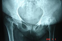
The bones in the immediate vicinity of the target organ of the radiation therapy are potentially at risk from osteoradionecrosis. For example, radiation from renal cell carcinoma can lead to osteoradionecrosis of the lumbar vertebrae . In gynecological tumors, the pelvic bones , in head and neck tumors, for example, the lower or upper jaw , in breast cancer, the ribs and bones of the shoulder girdle are at risk.
Osteoradionecrosis has a slow progression. In the course of time, they become more and more extensive and painful. If left untreated, they can lead to infection and pathological fractures . The damage caused by the osteoradionecrosis is usually irreversible.
In osteoradionecrosis in the lower jaw ( mandible ), in the after irradiation of tumors oropharynx may arise the danger of infection by the presence in this area is oral flora particularly high.
The exact pathogenesis of osteoradionecrosis is not yet fully understood. One theory assumes that microvascular thromboses , which are caused by destruction of the endothelium and inflammatory processes, lead to bone and tissue necrosis. The endothelium and the inflammatory processes are caused by free radicals that are generated by the ionizing radiation. There are also risk factors such as tobacco and alcohol abuse and surgical interventions. According to this theory, the administration of steroidal anti-inflammatory drugs before and after radiation therapy reduces the risk of osteoradionecrosis.
frequency
The incidence and likelihood of osteoradionecrosis depends on many factors. In addition to the dose, the size of the irradiated tissue volume, the type of bone ( long bones , flat bones , cancellous or compact bones, etc.) and the fractionation , the bone density , for example, also plays an important role. For example, the likelihood of osteoradionecrosis increases significantly in patients with osteoporosis .
The incidence of osteoradionecrosis within five years after radiation therapy is up to 11% after radiation therapy in the pelvic area (mainly for radiation of gynecological tumors) and between 5 and 10% on the lower jaw.
In the early days of radiation therapy, bone necrosis was relatively common. With the improvement of irradiation techniques, the incidence decreased significantly from the 1970s, but has increased slightly since the 1990s. A possible cause for this increase is the combination of chemotherapy and radiation therapy .
diagnosis
Osteoradionecrosis can be radiologically clearly seen as poorly demarcated dense bone debris. This allows them to be distinguished from bone metastases . In the case of mixed osteoplastic / osteolytic bone metastases, the differential diagnosis is much more difficult. A bone scintigraphy can help clarify the findings. A bone biopsy offers maximum diagnostic reliability .
Prophylaxis and therapy
The acute and chronic inflammatory processes of osteoradionecrosis are usually treated with steroidal anti-inflammatory drugs. If these anti-inflammatory drugs are given prophylactically before and immediately after radiation therapy, the risk of osteoradionecrosis can be reduced. In addition, the administration of pentoxifylline and an antioxidant treatment, for example with superoxide dismutase and tocopherol (vitamin E), is recommended.
In the jaw area, the risk of osteoradionecrosis can be significantly reduced by a thorough dental rehabilitation before starting radiation therapy.
The therapeutic benefit of hyperbaric oxygenation (HBO) in osteoradionecrosis is controversial. In addition to studies that describe a positive effect, there are studies that do not certify this treatment option to have a positive effect.
Medical history
In 1926, the American pathologist James Ewing was the first to observe bone changes as a result of radiation therapy, which he referred to as radiation osteitis (English : ' radiation ostitis ' or radio osteomyelitis ; today: osteoradionecrosis). For a long time it was assumed that the radiation causes a bacterial infection in the bone, which ultimately leads to radiation necrosis. It was not until 1983 that Robert E. Marx discovered that osteoradionecrosis is radiation-induced aseptic bone necrosis.
further reading
- AS Jacobson, D. Buchbinder et al .: Paradigm shifts in the management of osteoradionecrosis of the mandible. In: Oral Oncology . Volume 46, Number 11, November 2010, pp. 795-801, ISSN 1368-8375 . doi: 10.1016 / j.oraloncology.2010.08.007 . PMID 20843728 . (Review).
- DE Peterson, W. Doerr et al .: Osteoradionecrosis in cancer patients: the evidence base for treatment-dependent frequency, current management strategies, and future studies. In: Supportive care in cancer. Volume 18, Number 8, August 2010, pp. 1089-1098, ISSN 1433-7339 . doi: 10.1007 / s00520-010-0898-6 . PMID 20526784 . (Review).
- BR Chrcanovic, P. Reher et al: Osteoradionecrosis of the jaws - a current overview - part 1: Physiopathology and risk and predisposing factors. In: Oral and maxillofacial surgery. Volume 14, Number 1, March 2010, pp. 3-16, ISSN 1865-1569 . doi: 10.1007 / s10006-009-0198-9 . PMID 20119841 . (Review).
- BR Chrcanovic, P. Reher et al: Osteoradionecrosis of the jaws - a current overview - Part 2: dental management and therapeutic options for treatment. In: Oral and maxillofacial surgery. Volume 14, Number 2, June 2010, pp. 81-95, ISSN 1865-1569 . doi: 10.1007 / s10006-010-0205-1 . PMID 20145963 . (Review).
- P. Kouyoumdjian, O. Gille et al .: Cervical vertebral osteoradionecrosis: surgical management, complications and flap coverage - a case report and brief review of the literature. In: European spine journal. Volume 18 Suppl 2, July 2009, pp. 258-264, ISSN 1432-0932 . doi: 10.1007 / s00586-009-0950-8 . PMID 19340464 . PMC 2899554 (free full text). (Review).
- MJ Wahl: Osteoradionecrosis prevention myths. In: International Journal of Radiation Oncology - Biology - Physics . Volume 64, Number 3, March 2006, pp. 661-669, ISSN 0360-3016 . doi: 10.1016 / j.ijrobp.2005.10.021 . PMID 16458773 . (Review).
Individual evidence
- ↑ a b R.O. Ayorinde, CA Okolo: Concurrent femoral neck fractures following pelvic irradiation: a case report. In: Journal of medical case reports. Volume 3, 2009, pp. 9332, ISSN 1752-1947 . doi: 10.1186 / 1752-1947-3-9332 . PMID 20066055 . PMC 2804621 (free full text).
- ↑ a b c d e f g J. Freyschmidt: Skeletal diseases : clinical-radiological diagnosis and differential diagnosis. Edition 3, Springer, 2008, ISBN 3-540-45529-9 , p. 140. Limited preview in the Google book search
- ↑ J. Silvestre Rangil, FJ Silvestre: Clinico-therapeutic management of osteoradionecrosis: a literature review and update. (PDF; 463 kB). In: Medicina oral, patología oral y cirugía bucal. Volume 16, Number 7, November 2011, pp. E900 – e904, ISSN 1698-6946 . PMID 21743407 .
- ↑ a b S. Delanian, JL Lefaix: Current management for late normal tissue injury: radiation-induced fibrosis and necrosis. In: Seminars in Radiation Oncology . Volume 17, Number 2, April 2007, pp. 99-107, ISSN 1053-4296 . doi: 10.1016 / j.semradonc.2006.11.006 . PMID 17395040 . (Review).
- ↑ C. Madrid, M. Abarca, K. Bouferrache: Osteoradionecrosis: an update. In: Oral oncology. Volume 46, Number 6, June 2010, pp. 471-474, ISSN 1368-8375 . doi: 10.1016 / j.oraloncology.2010.03.017 . PMID 20457536 . (Review).
- ↑ J. Bahnsen, G. Hänsgen and others: Female basin. In: M. Wannenmacher, J. Debus, F. Wenz (Eds.): Radiation therapy. Springer, 2006, ISBN 3-540-22812-8 , p. 630. Limited preview in the Google book search
- ↑ D. Thönnessen, H. Hof et al: Head and neck tumors. In: M. Wannenmacher, J. Debus, F. Wenz (Eds.): Radiation therapy. Springer, 2006, ISBN 3-540-22812-8 , p. 412. limited preview in Google book search
- ↑ M. Wannenmacher, J. Debus, F. Wenz: General principles. In: M. Wannenmacher, J. Debus, F. Wenz (Eds.): Radiation therapy. Springer, 2006, ISBN 3-540-22812-8 , p. 7. limited preview in Google book search
- ↑ G. Laden: Hyperbaric oxygen therapy for radionecrosis: clear advice from confusing data. In: Journal of clinical oncology. Volume 23, Number 19, July 2005, p. 4465; author reply 4466-4465; author reply 4468, ISSN 0732-183X . doi: 10.1200 / JCO.2004.00.9829 . PMID 15994160 .
- ↑ RJ Shaw, J. Dhanda: Hyperbaric oxygen in the management of late radiation injury to the head and neck. Part I: treatment. In: British Journal of Oral & Maxillofacial Surgery . Volume 49, Number 1, January 2011, pp. 2-8, ISSN 1532-1940 . doi: 10.1016 / j.bjoms.2009.10.036 . PMID 20347191 . (Review).
- ↑ D. Annane, J. Depondt et al: Hyperbaric oxygen therapy for radionecrosis of the jaw: a randomized, placebo-controlled, double-blind trial from the ORN96 study group. In: Journal of clinical oncology . Volume 22, Number 24, December 2004, pp. 4893-4900, ISSN 0732-183X . doi: 10.1200 / JCO.2004.09.006 . PMID 15520052 .
- ↑ J. Ewing: osteitis Radiation. In: Acta Radiologica. Volume 6, 1926, pp. 399-412.
- ^ AS Jacobson, D. Buchbinder et al .: Paradigm shifts in the management of osteoradionecrosis of the mandible. In: Oral oncology. Volume 46, Number 11, November 2010, pp. 795-801, ISSN 1368-8375 . doi: 10.1016 / j.oraloncology.2010.08.007 . PMID 20843728 . (Review).
- ^ RE Marx: Osteoradionecrosis: a new concept of its pathophysiology. In: Journal of oral and maxillofacial surgery. Volume 41, Number 5, May 1983, pp. 283-288, ISSN 0278-2391 . PMID 6572704 .
- ↑ MM Baltensperger, GK Eyrich: Osteomyelitis of the Jaws. Springer, 2009, ISBN 3-540-28764-7 , p. 15. Restricted preview in the Google book search
