
tomography

The tomography or tomography (from ancient Greek τομή , tome , cut 'and γράφειν , graphein , write') is a imaging technique that provides the representation of an object in layers. The term covers various recording methods that determine the internal spatial structures of an object and can represent them in the form of sectional images . Synonymous terms are cross-sectional imaging and -shoot technique , the sectional images are also slice images or tomograms called.
A sectional image shows the internal structures as they would be after cutting the object or after cutting out a thin slice. One speaks here of an overlay-free representation of the corresponding object layer - in contrast to projection methods such as the usual X-ray examination, in which all structures that lie one behind the other in the beam path are superimposed. This difference is illustrated in the adjacent figure with two tomographic slice images (S 1 and S 2 ) and a projection image (P) of the same volume.
Tomographic methods can record a single slice or larger volumes, which can then be displayed as a series of parallel slice images, for example. Methods that record individual slices can also be used to record three-dimensional data sets by scanning the object in a series of parallel cross-sectional images.
Tomographic methods are particularly important in medical imaging , but some methods from geosciences , physics , paleontology or materials science can also be classified under tomographic methods.
Procedure in medicine
The following tomographic procedures are particularly important in medicine:
- the "classic" X-ray tomography ,
- ultrasound diagnostics ( sonography ),
- the computed tomography (CT),
- the magnetic resonance imaging (MRI, Magnetic Resonance Imaging),
- the positron emission tomography (PET),
- the single-photon emission computed tomography (SPECT),
- The optical coherence tomography (OCT), and
- the Electrical Impedance Tomography (EIT).
The basics of tomography in medicine in the form of "classic" X-ray tomography were developed by the radiologist Alessandro Vallebona in Genoa in 1930 .
The difference between the overlay-free display in medical tomography and a projection image is illustrated in the following figures. In projection methods such as radiography (normal X-ray examination), a shadow image is recorded on which several structures are superimposed when they lie one behind the other in the beam path. For example, in conventional x-rays, the soft tissues of the anterior and posterior chest wall and the bony structures of the thorax overlay the lung structures. This would make it more difficult to diagnose a lung tumor (e.g., lung cancer ). Each CT or MRT slice image of the thorax, on the other hand, shows only a 0.5 to 10 mm thick layer that is practically superimposed.
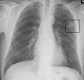
X-ray image (projection image ) of a small peripheral bronchial carcinoma in the left upper lobe
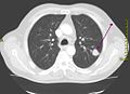
The same tumor on computed tomography (axial cross-section)

From the CT composite -Einzelschichten 3D image
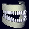
Reconstructed 3D tooth model from data from a DVT scan


Each pixel in a sectional image corresponds to a volume element ( voxel ) of the entire three-dimensional data set. The height of the voxel corresponds to the slice thickness. In post-processing, slice images ( multiplanar reconstruction , MPR), layered (sub-volume) maximum intensity projections (MIPs) or three-dimensional images of the examined object ( volume rendering ) can be calculated from the voxels .
Procedure in geosciences
In other areas of non-destructive testing , similar methods are used that work according to comparable mathematical inversion methods . These are:
- in geophysics e.g. B. geoelectrical , seismic and muon tomography as well as ground penetrating radar
- in geodesy , the laser scanning and methods of altimetry and gravimetry
Procedure in physics
- Quantum tomography , which enables the complete measurement of the quantum state of an object (e.g. its density matrix or its position and momentum distribution) using mathematical methods similar to those used in medicine
- Electron tomography, in which the individual sectional images (projections) are generated with the transmission electron microscope (TEM).
Other tomographic procedures
- Neutron Tomography : Applications in Paleontology and Materials Science
- Tomographic atom probe , atom-probe tomography (APT) or three-dimensional atom probe (3DAP): applications in materials science
- Photoacoustic Tomography (PAT): Applications in materials science and biomedical research
- Computed Tomography Imaging Spectrometer (CTIS): Process for the spectral recording of images
Individual evidence
- ↑ Ralf Habel, Michael Kudenov, Michael Wimmer: Practical Spectral Photography