Slit lamp
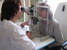
The slit lamp (also: slit lamp microscope ) is one of the most important ophthalmological examination devices with which the ophthalmologist or optician can inspect the eyes stereoscopically . It was the Swedish physician and Nobel Prize carrier Allvar Gullstrand introduced in ophthalmology 1911th
History of the slit lamp
For microscopic examination of the eye led Gullstrand 1910 by a Nernst a powered slit illumination. The industrial production of such slit lamps, albeit with equally bright incandescent light, initially took place at Carl Zeiss in Jena and Haag-Streit in Bern. The extensions developed there according to Goldmann, Henker, Koeppe, Vogt and others quickly made the slit lamp the most important examination device in ophthalmology. In 1930 Rudolf Thiel succeeded for the first time in photographing a segment of the eye that had been inspected with a slit lamp. The coupling of the slit lamp with a laser light source in the middle of the 20th century has enabled surgical measures since then and made the slit lamp the most versatile instrument in ophthalmology.
A historical model
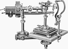
The picture on the left shows a complete slit lamp device, probably from 1924. The light source is a carbon arc lamp , which is attached to a tripod that can be freely pivoted in all directions and height-adjusted. Compared to the Nernst burning lamp, it provides a much brighter light, but the light rays, which can reach a color temperature of up to 5000 K , have to be cooled down using a water chamber (cooling cuvette). The cuvette is mounted on the so-called hangman arm (named after Otto Henker) on a sliding rider. In the beam path behind, also movable by means of a rider on the arm, the blind tube follows in the function of a lens hood . At the end of the pipe there are two Rekoss discs, with the help of which it is easy to change the pre-installed filters. At the end of the arm is the aplanatic aspherical lighting lens according to Allvar Gullstrand and the mirror device according to Leonard Koeppe. In the center of the picture, the actual corneal microscope according to Siegfried Czapski is shown with an objective revolver and height adjustment on a cross table; on the right in the picture the headrest with chin and forehead rest for the patient.
annotation
- ↑ Rekoss disk, named after the instrument maker Egbert Rekoss, who was a Hermann von Helmholtz institute mechanic in Königsberg.
Function and application
This optical device offers the examiner the possibility of directing a sharply delimited, slit-shaped light beam, the width of which can be changed, onto the eye. At the same time, he has the opportunity to look at it through a reflected light microscope . The magnification of the microscope is variable in most devices and usually ranges from 6 to 30 times.
Using various exposure methods (diffuse, direct, focal, indirect, regressive, lateral, etc.) and variable light slit widths, it is possible to inspect almost all the anterior, central and posterior sections of the eye right up to the retinal areas located far in the periphery . For some examinations, additional aids, such as a three-mirror contact lens , are necessary.
Most modern slit lamp are additionally equipped with an applanation tonometer according to Goldmann equipped, the measurement of intraocular pressure is used. A combination with digital cameras is also possible in order to document findings on film or photographically.
The production and quality of slit lamps is regulated in Germany by the Precision Mechanics and Optics Standards Committee (NAFuO) in DIN EN ISO 10939: 2007.
Web links
- Eilhard Koppenhöfer: From the side lighting to the slit lamp. A brief medical history. Kiel 2011, (PDF; 21.3 MB - worth reading).
literature
- Theodor Axenfeld (founder), Hans Pau (ed.): Textbook and atlas of ophthalmology. With the collaboration of Rudolf Sachsenweger a . a. 12th, completely revised edition. Gustav Fischer, Stuttgart a. a. 1980, ISBN 3-437-00255-4 .
- H. Slezak, P. Kenyeres: Slit lamp photography of the retinal rim and pars plana of the ciliary body. In: Albrecht von Graefes Archive for Clinical and Experimental Ophthalmology. Vol. 185, No. 4, 1972, ISSN 0065-6100 , pp. 269-274, doi: 10.1007 / BF00410757 .
Individual evidence
- ^ Alfred Vogt: Atlas of the slit lamp microscopy of the living eye. With instructions on the technique and methodology of the examination . Julius Springer, Berlin 1921.
- ↑ Eye examinations with the slit lamp . (PDF; 1.4 MB) Carl Zeiss Meditec, Jena 2006, p. 38 f., Dog.org; accessed on January 14, 2015.
- ↑ From the side lighting to the slit lamp. A brief medical history.
- ↑ Ophthalmic instruments - slit lamps, DIN EN ISO 10939.


