Epidermis (vertebrates)
The epidermis ( Greek epi "on", "above"; derma "skin") is the epidermis of vertebrates . As the outermost layer of the skin, it forms the actual protective cover against the environment .
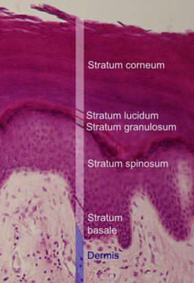
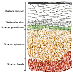
From the inside to the outside there are five layers of the epidermis: the basal layer ( stratum basale ), the prickly cell layer ( stratum spinosum ),
Granular layer ( stratum granulosum ), glossy layer ( stratum lucidum ) and horny layer ( stratum corneum ). The epidermis consists of 90 percent keratinocytes , which are held together by desmosomes . In the outer layers, it consists of keratinized squamous cells .
Stratum basale
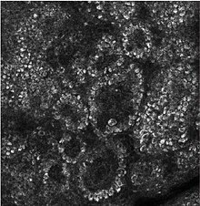
The stratum basale - the "basal cell layer" - serves as a single, innermost cell layer for the regeneration of the skin; Here is the cell division instead. One daughter cell begins its migration to the surface, the other remains and divides again. The supply of nutrients is still comparatively good here, although the epidermis itself does not contain any blood vessels. As with all epithelia, it is demarcated from the dermis below by a basement membrane , which runs largely flat in the field skin , but is strongly deformed in the inguinal skin by papillary bodies (bulges in the dermis), the density of which determines the structure of the dermal ridges .
Within the basal cell layer, there are special sensory cells for touch stimuli, the Merkel cells . Melanocytes , the pigment-forming cells, are also located here .
Stratum spinosum
In the stratum spinosum , also known as the “prickly cell layer”, the cells are connected to desmosomes by cytoplasmic extensions . This is where the step-by-step cornification process known as keratinization begins . Since the cells shrink during histological processing, they have a prickly appearance in the specimen. The stratum spinosum also contains defense cells of the lymphatic system , which are known as Langerhans cells .
Stratum basale and stratum spinosum are collectively referred to as stratum germinativum (synonymous: germ layer, regeneration layer).
Stratum granulosum
As the cornification progresses, the cells begin to break down in this “granular cell layer”, and they gradually transform into lifeless corneocytes. The outer shape gradually flattens out and the inside of the cell is more and more dominated by keratin granules . The cell nucleus is not as clearly delineated as in the stratum spinosum .
Stratum lucidum
The stratum lucidum , also known as the “glossy layer ”, is a layer of cells that looks very uniform under the microscope and is only found on the groin skin of the hands and feet. Its task is to represent a barrier against all forms of intruders into the skin. For the most part, it consists of an oily layer with minor differences in refraction (therefore it appears transparent). Here, keratohyalin granules liquefy to a semi-liquid, fat and protein-rich, acidophilic substance, elidin . In the skin of the field, the stratum lucidum is barely developed and can therefore only be recognized as a thin, differently colored cell strip under the almost unstructured stratum corneum . Here it forms the transition layer to the highly inhomogeneous granule cell layer.
Stratum corneum

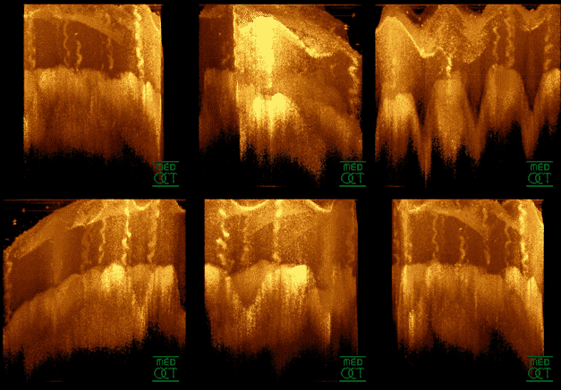
The transition to the stratum corneum , the outermost layer of the epidermis, occurs abruptly. The now completely cornified corneocytes now form the "horny cell layer" as "horny cells", which can be between 12 and 200 cell layers thick depending on the region. The cells of this epidermal layer have died and no longer contain any cell organelles . Fats between the cells and the horny cells form a water-repellent protective layer. In addition, special, species-dependent structures such as claws , nails , hooves , horns , horn sheaths , feathers and hair (the latter two only when the temperature is the same ), but also calluses and papillary patterns can form.
Stratum disjunctum
The stratum disjunctum is the uppermost layer of the cornea; due to the inclusion of air, it has a different contrast to the underlying material. Here the horny cells detach from each other by dissolving the contacts between the cells . Usually this process is invisible to the human eye. If the coordination of the exfoliation is disturbed, the corneocytes separate in larger associations. Aggregations of 500 or more cells are flakes of skin that are visible to the naked eye .
See also
Individual evidence
- ^ Rüdiger Wehner, Walter Gehring: Zoologie . 24., completely revised. Edition Thieme, Stuttgart 2007, ISBN 978-3-13-367424-9 , pp. 794 .


