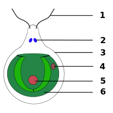
Eye spot

The eye spot (also called stigma ) is a sensory organically mostly photosynthetic , flagellated protist that serves for phototaxis . It lies outside or inside a chloroplast at the front pole or in the middle of the cell. They consist of photoreceptors and several lipid droplets , separated from one another by a basement membrane , in which carotenes are stored and therefore appear as bright orange-red pigment granules . Eye spots are the simplest and (as far as the number of individual wearers are concerned) the most common 'eyes' in nature. They are found in dinoflagellates , chlorophyta , yellow-green algae , brown algae , euglenoida , haptophyta and cryptophyceae . Signals transmitted by the photoreceptors of the eye-spots change the flapping pattern of the flagella and produce a phototactic response. The eye spot enables the cells to sense not only the intensity but also the direction of the light and react to it by swimming either in the direction of the light (positive phototaxis ) or away from the light (negative phototaxis). A related 'photophobic' reaction ('Photoshock') occurs when cells are briefly exposed to high light intensity. The cell stops, briefly swims backwards and then continues in another direction. The light perception mediated by eye spots thus helps the cells to find an environment with optimal light conditions for photosynthesis.
Microscopic structure
Under the light microscope , eye spots appear as dark, orange-reddish spots or stigmata. They get their color from carotenoid pigments, which contain pigment granules. The photoreceptors are located in the plasma membrane overlying the pigmented granules.
The Euglena eye spot includes the paraflagellar body that connects the eye spot to the flagellum . In electron microscopy the eye spot appears as a highly ordered lamellar structure.

In Chlamydomonas , the eye spot is part of the chloroplast and appears as a sandwich structure made up of several membranes. It is composed of the outer , inner and thylakoid membranes of the chloroplast, which enclose granules filled with carotenoids. The granulate stacks act as a retardation plate (more precisely quarter wave plate, λ / 4 plate) and reflect incident photons (light quanta) back to the photoreceptors above, while at the same time shielding the photoreceptors from light from other directions. The structure disintegrates during cell division and re-forms in the daughter cells in an asymmetrical manner to the cytoskeleton . This asymmetrical positioning of the eye spot in the cell is essential for correct phototaxis.
Eye spot proteins
The most important eye-spot proteins are the photoreceptor proteins that sense light. The photoreceptors found in unicellular organisms can be divided into two main groups: flavoproteins and rhodopsins ( English retinylidene proteins ). Flavoproteins are characterized in that they contain flavin molecules as chromophores (color carriers), while the retinylidene proteins contain retinal . The photoreceptor protein in Euglena is likely a flavoprotein. In contrast, the Chlamydomonas phototaxis is mediated by rhodopsins of the archaea type.
In addition to the photoreceptor proteins, eye spots contain a large number of structural, metabolic and signal proteins. In Chlamydomonas , the eyespot proteome consists of around 200 different proteins.
Photoreception and signal transmission
In Euglena a blue-light activated was as a photoreceptor adenylyl cyclase identified. The stimulation of this receptor protein leads to the formation of cyclic adenosine monophosphate (cAMP) as a second messenger (secondary messenger substance). The chemical signal transduction (signal transmission) ultimately triggers changes in the beat pattern of the flagella and in cell movement.
In Chlamydomonas the rhodopsins from archaea type contain a chromatophore with all- trans -Retinyliden, a photoisomerization to a 13- cis - isomer is received (i.e., under the influence of light that passes from the general retinylidene.. Trans - isomeric in the 13- cis -form over). This activates a photoreceptor channel, which leads to a change in the membrane potential and the cellular calcium ion concentration . The photoelectric signal transduction ultimately triggers changes in flagella movements and thus cell movements.
Strombidium
A special case of eye spots was found in the ciliate Strombidium tintinnodes syn. oculatum found: This uses green algae of the suborder Chlamydomonadina (order Chlamydomonadales ) as endosymbionts. However, these multiply faster than their host, so that the excess endosymbionts are broken down - except for stigmata, which collect in the front part ('apical') of the host and form an 'eye spot'. These stolen stigmata come from the chlorophyte prey and are therefore (by definition) kleptoplastids .
See also
Individual evidence
- ^ Rudolf Röttger: Dictionary of Protozoology In: Protozoological Monographs, Volume 2, 2001, p. 26, ISBN 3826585992
- ↑ a b P. Hegemann: Vision in microalgae . In: Planta . 203, No. 3, 1997, pp. 265-274. doi : 10.1007 / s004250050191 . PMID 9431675 .
- ↑ G. Kreimer: The green algal eyespot apparatus: A primordial visual system and more? . In: Current Genetics . 55, No. 1, 2009, pp. 19-43. doi : 10.1007 / s00294-008-0224-8 . PMID 19107486 .
- ↑ a b J. clouds: Euglena: the photoreceptor system for phototaxis . In: J Protozool . 24, No. 4, 1977, pp. 518-522. doi : 10.1111 / j.1550-7408.1977.tb01004.x . PMID 413913 .
- ↑ C. Dieckmann: Eyespot placement and assembly in the green alga Chlamydomonas . In: BioEssays . 25, No. 4, 2003, pp. 410-416. doi : 10.1002 / bies.10259 . PMID 12655648 .
- ↑ a b Suzuki T, Yamasaki K, Fujita S, Oda K, Iseki M, Yoshida K, Watanabe M, Daiyasu H, Toh H, Asamizu E, Tabata S, Miura K, Fukuzawa H, Nakamura S, Takahashi T: Archaeal- type rhodopsins in Chlamydomonas: model structure and intracellular localization . In: Biochem Biophys Res Commun . 301, No. 3, 2003, pp. 711-717. doi : 10.1016 / S0006-291X (02) 03079-6 . PMID 12565839 .
- ↑ Schmidt M, Gessner G, Luff M, Heiland I, Wagner V, Kaminski M, Geimer S, Eitzinger N, Reissenweber T, Voytsekh O, Fiedler M, Mittag M, Kreimer G: Proteomic analysis of the eyespot of Chlamydomonas reinhardtii provides novel insights into its components and tactic movements . In: Plant Cell . 18, No. 8, 2006, pp. 1908-1930. doi : 10.1105 / tpc.106.041749 . PMID 16798888 . PMC 1533972 (free full text).
- ↑ Iseki M, Matsunaga S, Murakami A, Ohno K, Shiga K, Yoshida K, Sugai M, Takahashi T, Hori T, Watanabe M: A blue-light-activated adenylyl cyclase mediates photoavoidance in Euglena gracilis . In: Nature . 415, No. 6875, 2002, pp. 1047-1051. doi : 10.1038 / 4151047a . PMID 11875575 .
- ↑ Strombidium oculatum Gruber, 1884 , on: World Register of Marine pecies (WoRMS)
- ↑ Taxon: Suborder Chlamydomonadina (alga)
- ↑ David JS Montagnes, Chris D. Lowe, Alex Poulton, Per R. Jonsson: Redescription of Strombidium oculatum Gruber 1884 (Ciliophora, Oligotrichia) , in: The Journal of Eukaryotic Microbiology , July 12, 2015, doi: 10.1111 / j.1550 -7408.2002.tb00379.x
- ^ E. Fauré-Fremiet: The Origin of the Metazoa and the Stigma of the Phytoflagellates , in: Journal of Cell Science 1958, pp. 3-99: pp. 123-129; PDF1 @paperity