Flagellum
Flagella ( Latin flagellum ) or flagella are thread-like structures on the surface of individual cells that are used for locomotion . They are fundamentally different in prokaryotes (living things without a cell nucleus ) and in eukaryotes (living things with a cell nucleus) in terms of structure and functionality:
- Prokaryotes have coiled protein threads outside of the cell membrane , which do not actively deform, are set in rotation by a motor at their end anchored in the cell and in this way - similar to a propeller - exert a thrust or pull .
- Eukaryotes, on the other hand, have filamentous protuberances of the cell enclosed by the cell membrane , inside of which there is a bundle of microtubules , and which cause movement by actively changing their shape.
These two completely different types of organelle are called both flagella and flagella , so these two names are used as synonyms . However, in order to take account of the different structure and the different functions of prokaryotic and eukaryotic flagella in terms of nomenclature, German-speaking authors have suggested the following language regulation:
“The flagella of the eukaryotic cells have a very uniform structure. [...] The term 'flagella' is reserved for the very differently organized locomotion organelles of the prokaryotes. "
In this article, this clear assignment of the terms “flagellum” for prokaryotes and “scourge” for eukaryotes is largely taken into account (difficulties arise, for example, with “flagellation”, see below). It should be noted, however, that this distinction is not made in other current textbooks.
The flagella of the prokaryotes
construction

Bacterial flagella are extracellular, helical threads (" filaments ") that are anchored in the cell membrane (or the cell membrane) and the cell wall via a "hook" with a motor complex . The flagella, including the hook and motor complex, are made entirely of proteins . The diameter of the filaments in most flagella is around 15–20 nm and they are hollow. Because of their small diameter, they can only be made visible with dark field microscopy and electron microscopy, not with normal light microscopy , but there are special dyeing processes that thicken them to such an extent that they are visible with light microscopy .
When the filaments are built up, the protein molecules ( flagellin ) are transported through the hollow channel of the flagella to the outer end and grown there. If there is a sufficiently large supply of flagellin in the cell, the build-up of a filament can happen very quickly.
The flagella of archaea are functionally similar to those of bacteria, but consist of different proteins and a different motor complex that is driven by ATP .
Flagellation types

According to the arrangement and number of flagella, different types of flagella are distinguished (in the order of descending swimming speed):
- holotrich : Numerous flagella are evenly distributed over the entire cell surface and encompass the entire body surface
- peritrich : many flagella are evenly distributed over the cell surface.
- polytrich-bipolar : The flagella are in two opposite groups at the cell poles. (also known as amphitrich ).
- polytrich-monopolar : The flagella stand in a group at one of the cell poles (also known as lophotrich ).
- monotrich : the cell has only one flagella.
- polar : The flagellum or the flagella are at one or both poles of the cell.
- lateral, lateral flagella : flagella stand laterally, not at the poles of the cell.
Lateral flagellation is often not associated with high swimming speed, but it has the advantage that the bacterium can more easily squeeze into obstacles such as highly viscous liquids or gaps between solids.
Way of movement
Due to their coiling, the flagella act like a propeller. The motor complex converts a difference in the concentration of protons between the two sides of the inner cell membrane into a rotating movement of the coiled filament sitting on a curved “hook” and thus follows a similar construction principle as the ATP synthase . The flagella mechanism is the only known truly rotating joint in all of biology. The rotational frequency is around 40–50 Hz.
The direction of the flagella rotation caused by the motor in combination with the winding direction of the flagella spiral determines whether a push or a pull is exerted on the bacterial body. The direction of rotation caused by the motor can be reversed in a very short time, so that thrust and pull can change quickly.
Usually the direction of rotation of the flagella is such that they slide. This means that in monopolar flagellated bacteria they are at the rear end. The bacterial body rotates (more slowly) in the opposite direction (preservation of the angular momentum ).
In bipolar flagellated bacteria, the flagella at both ends rotate in opposite directions. As a result, the flagella of the rear end have a pushing effect, the flagella of the front end are bent backwards and rotate around the front end of the bacterial body, thus increasing the thrust. If the direction of rotation of the flagella is reversed, the filaments fold over, the rear end of the bacterium becomes the front end and the front end becomes the rear end, the bacterium swims in the opposite direction.
The flagella of peritrich flagellated bacteria rotate in the same direction, usually so that they push. In doing so, they combine to form a backward-facing, coiled bundle, also known as a "scourge braid", which pushes the bacterium forward. If the direction of rotation of the flagella of peritrich flagellated bacteria is reversed, the individual flagella project radially from the bacterial body and their pulling effect on the bacterial body is canceled out on average, causing the bacterium to tumble in one place in random motion.
The reversal of the direction of rotation of the flagella and the associated change in the direction of movement play an important role in taxia (see for example chemotaxis ).
The flagella of the eukaryotes
construction

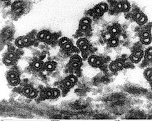
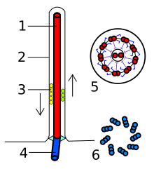
The flagella of the eukaryotes are thread-like structures that protrude from the body outwards into the surrounding medium and are surrounded by the cell membrane and filled with cytoplasm . Inside are microtubules in a special arrangement called 9 × 2 + 2; Nine double microtubules form a circle in the cross section with two single microtubules in the middle. The double microtubules each consist of a complete microtubule (A-tubule) and an incomplete (B-tubule). At the same height, about every 20 nm, there are pairs of protein arms on the A-tubule, which are called dynein arms. This microtubule arrangement is referred to as a 9 × 2 + 2 structure, the entire microtubule bundle as the axoneme . This structure is stabilized by various bridging proteins (especially nexin ). At the base of the flagella, where it merges into the cell body, there is a basal apparatus known as a blepharoplast or kinetosome, which is structurally similar to a centriol . It consists of nine triple microtubules in a circle (9 × 3 structure) that lies across a second, identically structured 9 × 3 structure. It is also often referred to as centriol. The eukaryote flagella are pointed at the free end. Their diameter is about 250-300 nm, their length a few micrometers to more than 150 µm.
Spermatozoa are a clear example of flagellated cells . The movement goes in a wave with constant amplitude from the base to the tip of the flagellum.
Together with the cilia , the flagella of the eukaryotes are also known as undulipodia .
Way of movement
According to the current state of knowledge, the change in shape required for hydrodynamic effectiveness comes about through mutually opposing sliding of the double fibrils. The energy for this should be provided by the dynein arms through hydrolytic cleavage of phosphate from ATP . The protein dynein , which forms the dynein arms, has ATPase activity. The sliding of the microtubules results in a change in the shape of the flagella.
The shape changes of the flagella are different depending on the type of flagella. They can consist of a wave (undulation) running over the flagellum in a plane or in the form of a helix with circular to elliptical movements, they can also consist of a flagellate stroke, whereby the flagellum curves in one direction and, so to speak, infiltrates the medium and strikes in the opposite direction, thus exerting a force. Flagella that move in the manner just described are called cilia . They are usually shorter than other flagella and arranged in greater density on the surface of the cells and tissues.
The result of the flagellum movement can be a movement of the individual, but it can also result in a movement of the adjacent medium or particles in the vicinity even when the individual is at rest. Examples of locomotion of the individual: freely moving ciliates , flagellates , spermatozoa . Examples of the movement of the adjacent medium or of particles: fixed ciliates, ciliated epithelium in the trachea of animals.
Variations, flagellation types
In some unicellular algae and protozoa are the flagella with many lateral short filaments, so-called Mastigonemen or fibrillation, and are occupied as Flimmergeißeln designated. The mastigonemes can appear in one row (stichonematic) or in two rows (pantonematic).
If a cell carries several flagella, one speaks of isocontal flagella, if these are similar, for example in green algae, different flagellated cells are referred to as heterocontal or anisocontal , with a long, forward-facing ciliated flagellum and a short one usually serving as the pulling flagellum smooth drag whip is directed backwards, so for example in the Heterokontae . In contrast to the flagellated forms, cells without a flagellum are called akont .
According to the place of insertion of the flagella, a distinction is made between acrokont (at the front end, pull flag), pleurokont (at the side) and opisthokont (at the rear end, push flag ).
Individual evidence
- ↑ Kleinig, Maier: Cell Biology , 4th Edition, 1999, p 151st
- ↑ For example B. in the German translation of the 3rd edition of Alberts' Essential Cell Biology (2005) the English original "flagella" translated with "flagella (flagella)" and because of this equation, the flagella of the eukaryotes is referred to in the following text as "flagella", cf. . ibid. p. 625 ff.
- ↑ Isokont in the Springer Lexicon of Biology
- ^ Heterokont in Springer Lexikon der Biologie
- ↑ Akont in Springer Encyclopedia of Biology


