Cell surface
The inside of a cell is separated from the outside by the cell membrane . The cell surface is the contact area where a cell comes into contact with the outside world. The cell surface is also the part of a cell that determines how other cells, blood components such as antibodies , complement proteins , hormones or nutrients interact with a given cell.
The cell surfaces are characterized by proteins , glycoproteins , proteoglycans , glycolipids and membrane lipids . One can easily distinguish whether a component should have an external or internal effect.
Outward effect
- Histocompatibility antigens are cell surface complexes with which a cell shows the immune system that it belongs to the same individual and which cell type it belongs to.
- Antigen receptors on cells indicate that they are lymphocytes (cells of the immune system).
- Mannose receptors can be found on phagocytes that eat and dispose of aging cells.
It is not by chance that these cells, which have an external effect, belong to the immune system: a hormone-producing cell, whose function is also directed towards the outside, cannot be seen from the outside that hormone is stored inside, just waiting to be released. From the outside it is also not possible to tell which hormone. In immune cells , the recognition of the outside is already present as a function through proteins in the cell membrane and can be determined with the help of suitable analyzes ( monoclonal antibodies against the recognition structures).
- Almost all cells are in cell clusters and are linked to their neighboring cells by tight junctions . In desmosomes , keratin threads lie beneath the cell membrane , which are connected through the cell membrane to cadherins on the cell surface. Cadherins bind to the cadherins of neighboring cells, creating a seal that is impenetrable for cells and blood components.
- Muscle cells can only trigger movement in the cell structure. To do this, they have to bind their contractions to those of other cells and to fixed holding points (tendons), which requires characteristic proteins on the cell surface.
- During cell adherence , cells come into temporary contact. The proteins of the integrin family in particular mediate cell adherence by reacting with binding partners on other cells. White blood cells therefore often do not swim freely in the blood, but instead "roll" on the blood vessel wall: on the one hand, they are driven by the bloodstream, on the other hand, they attach themselves briefly to other cells and test their surface signals. In the event of an inflammation, messenger substances ( cytokines ) are sent out from the inflammation focus , which lead to changes in the surface proteins on the cells of the vessel wall. In this case, the immune cells no longer "roll", but attach themselves firmly and push themselves through the wall between cells in order to get attracted by the messenger substances to the inflammation focus.
Inward effect
The molecules with which it absorbs nutrients are relevant for every cell. Sugars such as glucose , amino acids or ions are all kept away from the cell interior by the cell membrane barrier and require transport systems to get into the cell. Every cell therefore needs glucose transporter proteins, transporters for essential amino acids that a human cell cannot produce itself, or ion channels. The characteristic functions of a cell type are expressed in a special protein program that is also used on the cell surface:
Nerve cells , for example, are characterized by the fact that they release neurotransmitters , but can also bind them. The possibility of being able to spill can not be seen on the surface, but you can see the glutamate , GABA , dopamine , noradrenaline , serotonin receptors , as well as those for all other neurotransmitters on nerve cell surfaces. The ion channels of neurons are also visible from the outside.
Cells of the hormone release cascades have receptors for release hormones . Since hormones swim in the blood, they could bind to any cell; however, a functional bond only takes place where a cell with the hormone receptor can determine the existence of a hormone in the fluid around the cell and reacts to the binding of the hormone to the receptor with an internal signal.
Cells that can reabsorb the water from the primary urine in the kidney have water transporters ( aquaporins ), the presence of which on the cell surface is regulated by hormone signals. These signals come from cells that can react to different levels of salt in the blood fluid (receptors for osmolarity ). Before such a reaction from the brain's osmolarity receptor reaches the nephrons , vasopressin must first be released into the blood in the pituitary gland , which is recognized by receptors in adrenal cells and there triggers the formation and release of aldosterone, which ultimately causes water retention in the adrenal gland .
Cells in which not just one cell but the organism itself comes into contact with the outside world have a special function: lung tissue, intestinal surface, skin, mucous membranes of the mouth and the genital organs. Here, the tissue surface is often still covered by a layer of mucus, which consists of proteoglycans. The proteoglycans were called mucins after the names mucous membrane and mucus (Latin) mucus . They can store a lot of water, which creates a moist layer on the cells, e.g. B. separates from the air. In direct contact with the air, the surfaces would dry out and the cell underneath would die. The formation of mucins is a characteristic feature of mucosa cells. In the skin, dead skin cells protect the underlying skin cells from drying out.
The view of cells
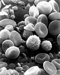
Cells are so small that the properties of their surface can only be seen with the help of an electron microscope ( scanning electron microscope ). On the left side, white blood cells are visible, which can be recognized mainly by the "rippled" surface. The red blood cells , the erythrocytes, can be recognized by their smooth surface and their concave / convex shapes (small bodies with different shapes are the platelets ). In the lower picture on the left you can see how a leukocyte torments its way through a blood vessel endothelium that has previously become adherent. On the right side you can see different views of the lung epithelium , from the lumen (above) and across the membrane (below). Depending on the type, the cells are equipped with long villi, short or not with villi. The different electron density and granularity in the picture below indicate the different functions of the epithelial cell types, which cannot be seen from the surface of the cell.
See also
literature
- Bruce Alberts : Textbook of molecular cell biology ("Essential cell biology"). 3rd edition Wiley-VCH, Weinheim 2005, ISBN 3-527-31160-2 .
- Charles Janeway : Immunology ("Immunobiology"). 5th edition. Spektrum Akad. Verlag, Heidelberg 2002, ISBN 3-8274-1079-7 .
- Bernhard Kleine: Hormones and the hormonal system. An endocrinology for life scientists . Springer Verlag, Berlin 2007, ISBN 978-3-540-37702-3 .
- Ivan M. Roitt et al. a .: Immunology ("Immunology"). 3. rework. Edition Thieme, Stuttgart 1995, ISBN 3-13-702103-0 .


