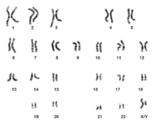
Karyogram

A karyogram ( Greek ϰάρυον káryon 'nut, fruit core' and γράμμα grámma 'written') is the orderly representation of all chromosomes in a cell . It is classified according to morphological aspects such as size, centromeric position and band pattern .
Karyograms are created to determine the karyotype , i.e. the chromosome makeup of an individual . The karyotype, in turn, can be used, for example, to compare the chromosome endowments of different species (e.g. humans and chimpanzees) or, in human genetics , to determine the cause of hereditary diseases.
Typical preparation of a karyogram in medicine
To create karyograms first chromosome preparations are made in which metaphase - chromosomes are adjacent. For this purpose, blood is taken from the patient, from which the lymphocytes are isolated ( erythrocytes are not suitable due to the lack of a cell nucleus ). They are cultured for three days in a special nutrient medium that contains a substance that stimulates cell division ( phytohemagglutinin ). The division of the nucleus (mitosis) is then interrupted in the metaphase by adding the mitosis inhibitor colchicine for two hours , since colchicine inhibits the development of the spindle apparatus .
In the metaphase of mitosis, the chromosomes are in a strongly condensed form and can therefore be easily recognized with a light microscope . The lymphocytes are sedimented in a pellet by centrifugation . The supernatant solution is aspirated and the lymphocytes are dissolved in a hypotonic solution (distilled water or 0.075 molar potassium chloride solution). The hypotonic solution causes the cells to swell and are then fixed with a methanol - glacial acetic acid mixture in a ratio of 3: 1 . In the next step, a drop of the cell suspension is dropped onto a slide using a pipette . When hit, the cells burst open. Since those cells that are in the metaphase do not have a nuclear membrane , the metaphase chromosomes usually come to lie next to each other on the slide.
They are stained with a suitable method (e.g. Giemsa staining , FISH test ). While photographs of the chromosomes used to be made under 1000x magnification and these were individually cut out by hand and sorted according to size, centromere region and band pattern , today this is done using image processing software on the computer.
The total costs of the medical creation of a human karyogram amounted to about 350 € in 2014.
Human karyogram
The human chromosome set consists of 22 autosomes ( homologous chromosome pairs) and one gonosome pair (sex-determining chromosome pair). The appearance of the entire set of chromosomes, the karyotype , can be described by producing a karyogram in a person with 46, XX (44 autosomes and 2 identical gonosomes in a woman) or 46 XY (44 autosomes and 2 different gonosomes in a man) . In normal human body cells there are two chromosomes, there is a double ( diploid ) set of chromosomes (2n = 46 chromosomes). The sorting of the chromosomes in the karyogram is done according to the criteria of the size of the chromosome and the position of the centromere, into 7 groups (A – G). Depending on the position of the centromere, one speaks of acrocentric (near the end), submetacentric (between middle and end) and metacentric (in the middle of the chromosome). Due to morphological properties, the X chromosome is assigned to group C and the Y chromosome to group G. Schematic representations of average chromosomes , the idiograms, serve to compare the assignment made .
| group | number | associated chromosomes | features |
|---|---|---|---|
| A. | 3 | 1-3 | Large, meta- or sub-metacentric |
| B. | 2 | 4 + 5 | Large, submetacentric |
| C. | 7th | 6-12 + X | Medium, meta- or sub-metacentric |
| D. | 3 | 13-15 | Medium, acrocentric |
| E. | 3 | 16-18 | Short, submetacentric |
| F. | 2 | 19-20 | Short, metacentric |
| G | 2 | 21 + 22 + Y | Very short, acrocentric |
All living beings that belong to the same species have a specific number of chromosomes in their cells, which is usually an even number, with their subspecies often having a multiple (2, 3 times) this basic number of chromosomes . Organisms closely related to one another often have the same or a similar number of chromosomes. However, an identical or similar set of chromosomes is not a clear indication of a relationship between different living beings. There is no direct correlation between the number of chromosomes and the level of organization of living beings (e.g. human 46 chromosomes vs. potato 48 chromosomes). However, there is a direct correlation with related species in terms of the number and size of chromosomes. The higher the number of chromosomes, the smaller the size of the individual chromosomes.
Karyogram to identify chromosomal abnormalities
In human genetics, the aim of a karyogram is to identify possible genetic risks and hereditary diseases of the family in advance or to exclude them. Furthermore, the execution of a karyogram is mainly used to determine gender. Hereditary diseases are caused by defective genes and chromosomal abnormalities , which are inherited. These changes in the chromosomes are caused by mutations .
There can be 3 different types of mutations. On the one hand, the mutation of individual genes, called gene mutation (e.g. phenylketonuria ). On the other hand, the mutation of the chromosome structure, which is referred to as chromosome or structure mutation (e.g. translocation trisomy 21 ). There is also the mutation of the chromosome number, called genome mutation . This is divided into aneuploidy , in which one or more chromosomes are increased or decreased (e.g. Down syndrome ), and euploidy , in which the entire set of chromosomes is increased or decreased (e.g. cultivated wheat is hexaploid).
In the context of biological research, the production of a karyogram is used to create the karyotype, to describe a certain species (e.g. animal or plant) or to characterize special cell cultures or tumor cells.
swell
- Werner Buselmaier and Gholamali Tariverdian: Human Genetics . Springer-Verlag, Berlin, Heidelberg, New York 1991, ISBN 3-540-54095-4 .
- Werner Buselmaier: Biology for Physicians . Springer-Verlag, Berlin, Heidelberg 2012, ISBN 978-3-642-00451-3 .
- Neil A. Campell: Biology . 2nd Edition. Spektrum Verlag, Heidelberg 1997, ISBN 3-8274-0032-5 .
- R. Hagemann, T. Börner , F. Siegemund, General Genetics. With 71 tables. 4th edition Heidelberg [u. a.]: Spektrum, Akad. Verl. (1999) Online
- Meinhard and Moisl: Biology 1, Genetics, Metabolism, Ecology . 1st edition. Stark-Verlag, 1995, ISBN 978-3-89449-201-4 .
Web links
Individual evidence
- ↑ a b c d Werner Buselmaier, Biology for Physicians; Page 139–153
- ↑ a b Neil A. Campell, Biology; Page 250
- ↑ a b c Meinhard / Moisl, Biology 1 - Genetics, Metabolism, Ecology; Page 3–6
- ↑ Meinhard / Moisl, Biology 1 - Genetics, Metabolism, Ecology; Page 5 Tab. 1
- ↑ http://www.ckjh.de/resources/bc-pnd-2.doc accessed on January 11, 2013
- ↑ a b Meinhard / Moisl, Biology 1 - Genetics, Metabolism, Ecology; Page 36
- ↑ http://www.artikel32.com/biologie/1/karyogramme.php accessed on February 15, 2013


