
Mitosis
As mitosis ( Greek. Μίτος mitos , thread ') or mitosis (Greek κάρυον. Karyon ' core ', κίνησις kinesis , movement'), also indirect nuclear division , the division of the is cell nucleus refers to arise in the two daughter nuclei with the same genetic information. It takes place in cells of eukaryotic organisms - prokaryotes have no nucleus - and usually precedes a division of the entire cell, from which two daughter cells emerge.

In the cell cycle of dividing cells of eukaryotes, nucleus division and cell division are linked to one another. Mitosis and cytokinesis are also referred to together as mitosis or M-phase . During the interphase between successive mitoses, the DNA molecule of a chromosome is duplicated ( replication ), after which each chromosome consists of two identical sister chromatids . During mitosis, these chromatids are then separated and divided so that each daughter nucleus receives an identical half as a daughter chromosome. This means that an identical copy of the entire chromosomal genome of the mother cell can be passed on to two daughter cells .
In mitosis there is no change in the set of chromosomes , the degree of ploidy remains the same. Was the starting cell haploid , including the nuclei of the daughter cells are haploid. Was the starting cell diploid , so the nuclei of the daughter cells are diploid.
Meiosis is to be differentiated from mitosis with a fundamentally different way of nuclear division, in which the sister chromatids are not separated in the reduction division, but are assigned together to a daughter nucleus. It is integrated into the generation cycle and leads to a reduction in the number of chromosomes and genetically different daughter cells.
history
The Tübingen botanist Hugo von Mohl first observed cell division under the microscope in 1835 in the green alga Cladophora glomerata , then also in land plants. In 1841 , Robert Remak first described the forms of division of animal cells in embryonic blood cells, and later the division of the fertilized egg cell in chickens with the development of three different cotyledons . He emphasized the importance of the phenomenon of cell division for the formation of new cells and suggested that the new cell nuclei are also formed by division. In the following years other cell researchers saw division processes in the cells of many plants and animals. Hugo von Mohl made a discovery that was important for understanding life processes in retrospect. The Berlin doctor Rudolf Virchow summed it up in 1855 in the expression Omnis cellula e cellula or
"Where a cell arises, a cell must have preceded it [...]."

However, there were still unclear ideas about the fine structure of the cell building blocks known at the time and their function. This particularly affected the nucleus and its role in division. It was only with the advancement of microscopes and staining techniques in the second half of the 19th century that researchers were able to gain new knowledge. As described 1873 Giessen zoologist Anton Schneider in the flatworm Mesostoma ehrenbergii ( "Glass Strudelwurm") the proceeding changes of the core of the division as well as a rosette-shaped arrangement thickened strands in "equatorial".
The Bonn botanist Eduard Strasburger also noticed stages of division in a preparation of dividing cells in 1874 with nuclear spindles instead of a normal cell nucleus in which elongated, curved or unwound thread structures were visible. The Kiel anatomist Walther Flemming called their substance chromatin because of their strong stainability and referred to the entire process of nuclear division in 1879 as “mitosis” (from the Greek word for “thread”). Previously, he had found in the cells of the fire salamander that each thread separates into two parallel ones, that the new nuclei arise from the full half of a spindle - and that nothing remains. The Berlin anatomist Wilhelm Waldeyer suggested the designation chromosomes in 1888 . A closer microscopic examination revealed that each chromosome consists of two identical halves, the chromatids . These lie close together, but are only connected to one another at one point, the centromere .
Chromosomes were not only discovered in plant and animal cells, but also in some (eukaryotic) unicellular cells. In the course of time it was found that every plant and animal species has a specific number of chromosomes in all body cells. The number is between two chromosomes in the horse roundworm ( Ascaris megalocephala univalens ) and a few hundred in some plants.
Function of mitosis
Mitosis makes it possible to split up the genetic information contained in the chromosomes in such a way that two daughter cell nuclei receive the same genetic information again . For this, the genetic material in the nucleus of a mother cell must have been doubled beforehand - during the preceding interphase of the cell cycle . Each chromosome, which after nuclear division initially consists of one chromatid, has two identical sister chromatids after duplication, which are connected at the centromere. In the mitotic phases, these are compressed, attached, arranged, each separated and each moved apart, so that two spatially different - but identical in terms of number and type of chromosomes - are created, between which the nucleus is then divided.

In unicellular eukaryotes , karyokinesis, together with cytokinesis, forms the basis for their reproduction when the cell divides after mitosis. In some of these protists , mitosis takes place in a similar way to the multicellular eukaryotes as open mitosis , i.e. the nuclear envelope is temporarily dismantled. However, the nuclear envelope is retained in various other protists, so that a closed mitosis takes place.
In multicellular eukaryotes, mitosis is the prerequisite for the formation of a new cell nucleus and usually - with a few exceptions - also for the formation of new cells . In multicellular organisms such as humans, cells no longer divide in the course of their development in all developed cell lines. Nerve cells and muscle cells no longer multiply after differentiation has been completed. These cells leave the division cycle post-mitotic and enter the so-called G0 phase , so that the DNA is not replicated in the first place (see cell cycle ). Mature red blood cells in humans can no longer divide because they then lack their cell nucleus and mitosis cannot be initiated. Epithelial cells in the intestine and in the epidermis, on the other hand, multiply much more frequently than the average and thus renew the inner and outer surfaces of the body.
In human cells, the actual nucleus division usually takes about an hour; the interphase of the cell cycle between the mitotic phases of continuously dividing cells lasts significantly longer, depending on the cell type, about 12-24 hours. In other organisms, the mitotic duration can be longer, such as with the broad bean with about two hours, or shorter, as with the fruit fly , where it is often only 9 minutes long.
Mitosis can be stimulated by various so-called mitogens . The process of nuclear division is then initiated by the mitosis-promoting factor (MPF), the protein compound of cyclin B with a kinase that is dependent on it ( CDK 1 ).
Delimitation of meiosis
A special type of nucleus division must be distinguished from mitosis, in which the chromosome set is reduced and no identical daughter nuclei are created. It occurs as meiosis or meiotic division in the formation of germ cells for sexual reproduction and can create four haploid cells from a diploid starting cell in two division steps . In the first step ( reduction division ) the set of chromosomes is halved, while the second division ( equation division ) roughly corresponds to the process of mitosis.
Forms of mitosis
Mitoses in the cells of eukaryotic organisms follow a similar process scheme, but not all in the same form. In this way, it is possible to differentiate between open and closed mitoses, and as an intermediate form with partial breakdown or breakthroughs of the nuclear envelope, a half-open mitosis can be distinguished according to whether the nuclear membrane surrounding the karyoplasm with the chromosomes is broken down during karyogenesis . In addition, with regard to the design of the spindle apparatus, approximately axially symmetrical forms as "orthomitosis" ( centered ) can be distinguished from others with eccentric spindles, which are referred to as "pleuromitosis" ( laterally displaced ). With reference to a preserved nuclear envelope, it is also possible to differentiate between intranuclear versus extranuclear forms based on the position of the spindle poles .

open orthomitosis

half-open orthomitosis

semi-open pleuromitosis
closed orthomitosis
closed intranuclear pleuromitosis

closed extranuclear pleuromitosis


A cell nucleus division only takes place in cells of living beings in the domain of the eukaryotes ( Eukaryota ), because those of the bacteria ( Bacteria ) and the Archaea ( Archaea ) do not have a nucleus. The eukaryotic creatures are classified differently taxonomically and summarized in different groups, supergroups or supergroups. A mitosis of the closed form is found within each of the supergroups , a mitosis of the open form as well, with the exception of Excavata , which only show closed mitoses.
- An open Orthomitose is typical of mammals as other Metazoa , and terrestrial plants ; but it also occurs with some protists .
- A half-open orthomitosis occurs in different variants in Euamoeba and in green flagellates such as Raphidophyta or Volvox, for example .
- A semi-open Pleuromitose is typical of most Apicomplexa , such as Plasmodium .
- A closed orthomitosis is found in diatoms , in ciliate animals , in some microsporidia , unicellular yeasts and also in multicellular fungi .
- A closed intranuclear pleuromitosis is typical for kinetoplastids , for example trypanosomes , for oxymonadids, foraminifera , radiation animals and some green flagellates.
- A closed extranuclear Pleuromitose occurs in trichomonads and dinoflagellates on.
Stages of mitosis

Overview
Mitosis is divided into several phases, which flow into one another. While in the classical German literature four main phases of mitosis are often distinguished, whereby the prophase is followed by the metaphase, especially in the English-language literature the prometaphase is regarded as an independent phase in between, with which five phases of mitosis are separated from each other. In this prometa phase, the nuclear envelope breaks down for open mitosis in cells of animals and plants.
- In the prophase of the animal cell, the two centrosomes separate and migrate to opposite poles of the cell. The centrosomes act as microtubules -organisierende Centers (English microtubule organ ising center , MTOC) and are sorted starting points for the formation of the mitotic spindle. In higher plants, other cell components take on the task than MTOC, because their cells do not have centrosomes. The chromosomes condense , are thus visible under a light microscope, and can only now be seen in the often represented X-shape (during the interphase they are stretched out up to several centimeters long, as thin, thread-like structures). Since the chromosomes have already been doubled in the interphase, they each consist of two identical sister chromatids that are still connected at the centromere . The end of the prophase is reached when the nuclear envelope fragments (English-language literature) or when the condensation of the chromosomes is complete (classic German literature).
- In the prometa phase , the nuclear envelope disintegrates and the spindle fibers of the spindle apparatus penetrate from both poles into the area of the now envelope- free caryoplasm. The kinetochore microtubules, which start in the area of the centromere, are differentiated from the star-shaped astral and the overlapping, connecting polar ones . The chromosomes can now be moved, aligned and arranged by means of the adhering microtubules.
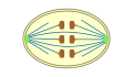
- In the metaphase , the highly condensed metaphase chromosomes are aligned by the microtubules as spindle fibers between the spindle poles in the equatorial plane . The metaphase is complete when all chromosomes have arrived in this metaphase plate , lined up and their kinetochores are connected to microtubules from both poles.
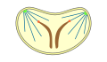
- In the anaphase , the two chromatids of a chromosome are separated and pulled apart along the spindle fibers, centromere first, in the opposite direction to the spindle poles. In this way, a complete set of chromatids or daughter chromosomes collects at each pole. This creates the basis for the two daughter cores. The anaphase is considered to have ended when the chromosomes of the two future daughter nuclei no longer move apart.
- The last phase of mitosis is called telophase . It seamlessly follows the preceding anaphase. The kinetochore fibers depolymerize, the nuclear envelope is rebuilt and the chromosomes decondense. After the decondensation is complete, the genes can be read again and the nucleus has its working form again.
In most cases, the telophase is followed by cytokinesis , with which the daughter nuclei can then be assigned to two daughter cells. However, this cell division is not part of mitosis.
Prophase

Following the interphase and the almost complete replication of the DNA, the previously loosely packed chromatin condenses , making the chromosomes recognizable as thread-like structures under a light microscope. The initially long, thin chromosomes each consist of a pair of chromatids that are held together at the central centromere. The chromatids fold and condense increasingly. In this compressed form, the DNA can no longer be read, a transcription of genes is impossible and the coded information can no longer be expressed . Therefore, in the prophase, the nucleoli dissolve as visible nuclear bodies, because the production of the ribosome components can no longer take place because of the chromosome compression.

Chromosome condensation
During the interphase, the chromatin is decondensed in the cell nucleus, the continuous DNA double strand of a chromosome is only loosely surrounded by packaging proteins in many places and is therefore accessible. At the beginning of the prophase, the chromatin threads condense and shorten through binding of condensins, increasingly through folding and multiple turns in loops, spirals and double coils. Their high degree of spiraling creates structures that are visible with a light microscope, the nuclear loops or chromatids of a chromosome. These are new structures in that they represent a more compact form of chromatin threads suitable for transport. In this state, the DNA segment of a gene is also inaccessible and cannot be expressed in this way.
In the prophase, every chromosome shows a longitudinal gap because it consists of two chromatids, each with a replicated DNA copy. The chromatids are held together at at least one point of constriction, the centromere.
Spindle fiber formation
In animal cells, two centrosomes (each with a pair of centrioles ) were formed by doubling during the interphase . They now migrate to opposite sides of the core and thus form the poles of the spindle. The structure of the spindle apparatus from microtubules is organized with the centrosomes . These represent the spindle fibers and are built up from tubulin subunits by polymerisation; they can also depolymerize again - just like other microtubules of the cytoskeleton when the cell becomes rounded. First of all, spindle fibers emanating from the centrosomes in a star shape are formed, which is also called an aster or astral microtubules .
For the microtubule-organizing centers (MTOC), the centrioles themselves are less important than the factors associated with them in the (pericentriolar) environment of a centrosome (after selective laser-surgical destruction of the centrioles, the functionality of the core spindle can remain unaffected). Plant cells manage without centrioles or centrosomes, where other structures take on the task of organizing microtubules as elements of the spindle apparatus. The spindle pole bodies in the cells of mushroom mushrooms also have no centrioles.
Prometaphase

In open mitosis, the nuclear envelope is temporarily broken down. This begins in the prometaphase through phosphorylation of the lamines , which are no longer attached to the inner membrane side of the double nuclear membrane as stabilizing intermediate filaments . The centrosomes, which are pushed further apart towards opposite poles, then form the starting point for spindle fibers. The sprouting spindle penetrates into the nucleoplasm from both poles , creating connections between the poles through overlapping, called polar microtubules . Three-layer kinetochores are formed on the centromeres of the chromosomes, to which so-called kinetochore microtubules attach. These are responsible for the transport of a chromosome - the chromatids that are only separated later - and are arranged parallel to the pile fibers.

Disintegration of the nuclear envelope
In animal cells, the prometaphase begins with the dissolution of the nuclear envelope . The fragments resulting from this decay can hardly be distinguished from parts of the endoplasmic reticulum .
In a number of eukaryotic unicellular organisms ( protozoa ), the nuclear envelope remains intact during the process of nuclear division and provides attachment points for the nuclear spindles. In trichomonads and some dinoflagellates , the centrioles in the cytoplasm are outside the preserved nuclear envelope; the two half-spindles of the extranuclear spindle apparatus come into contact with the chromosomes via the nuclear envelope.
Completion of the spindle apparatus
The star fibers or astral microtubules emanating from the centrosomes make contact with other elements of the cytoskeleton . Overlapping microtubules from one pole of the cell to the other, pole fibers or polar microtubules , also arise . So-called kinetochores are located on the centromeres of the chromosomes . As specific multilayered protein structures, they serve to bind tubulin and lead to the polymerization of microtubules , which form as kinetochore microtubules in the direction of the poles. These enable the movement and alignment of a chromosome as well as the subsequent separation of its chromatids in the area of the centromere.
Metaphase

The metaphase is the second phase of mitosis if the prometaphase is not viewed as a separate phase.
The chromosomes are now almost as short as possible. They are transported by pulling and pushing the spindle apparatus and are thus arranged in the equatorial plane at approximately the same distance from the two spindle poles in between . The chromosomes are thus next to each other in a starting position from which the sister chromatids can then be pulled apart. However, this only happens after all of their kinetochores are connected to microtubules.

Metaphase plate
The arrangement of the chromosomes in the equatorial plane at approximately the same distance from the spindle poles is also known as the metaphase plate . Microscopic images of this phase are used to visually identify individual chromosomes of a chromosome set in order to determine the karyotype .
A mitosis checkpoint also falls in this phase : only after the attachment of microtubules on the part of both poles of the bipolar spindle can the bond between the chromatids (by cohesins ) be broken . The metaphase changes into the anaphase when the sister chromatids of the chromosomes separate at the centromer site; then these migrate as daughter chromosomes, which now only consist of a chromatid, to the opposite poles.
Anaphase

The two chromatids of a chromosome are separated from each other and moved in different directions. The sister chromatids thus become daughter chromosomes (single chromatid chromosomes), which are transported along the spindle fibers to the opposite poles of the spindle. This shortens the kinetochore fibers. Meanwhile, the microtubules of the pile fibers can elongate, causing the poles to move apart.

Chromatid migration
The kinetochormicrotubules are roughly parallel to the pole fibers. According to recent research, it is assumed that the pulling forces from the polar directions are not decisive for the drifting apart of the chromatids, but rather motor proteins on the kinetochores, which migrate along the microtubule filaments in the direction of the centrosomes. This mechanism then follows a principle according to which the dynein or kinesin proteins also pull along a microtubule. The chromatids are slowly pulled apart from their central position in the equatorial plane towards the poles.
Anaphase I and Anaphase II
In the anaphase, a distinction can be made between the moving apart of the chromosomes - as anaphase I - and the moving apart of the spindle poles - as anaphase II.
Initiation of cell division
The simultaneous lengthening of the polar microtubules has the effect that the two polar regions that have formed in the cell are pushed further away from each other and so there are better conditions for cytokinesis. Later cell division can already be prepared in this phase of the nucleus division, also through interactions with actin filaments in the cortical part of the cytoskeleton below the cell membrane . The subsequent telophase, with which the cell nucleus division is completed, begins when the chromosomes arrive at both poles.
Telophase


When the daughter chromosomes finally reach the spindle poles, the increasingly shortened kinetochore fibers largely depolymerize. The polar fibers can initially lengthen even further until the poles have reached their maximum distance, then the spindle apparatus dissolves. The nuclear envelope of the daughter nuclei is now largely built up from fragments of the old nuclear membrane. The chromosomes decondense again. The nucleoli also reappear as corpuscles in the respective nucleus.
The division of the cytoplasm and thus the cell is described by cytokinesis.
Mitotic phase in the cell cycle
Cytokinesis

In most cases, after karyogenesis is complete, cytokinesis causes the cell to divide. In a cell cycle , mitosis and cell division are coupled in the mitotic phase .

In dividing animal cells, a contractile ring of actin fibers is formed during the telophase or already during the anaphase , which is narrowed together with myosin until the constrictions of the cell membrane fuse and daughter cells separated by this division groove are separated from each other.
In dividing plant cells, a special microtubule structure is formed in the equatorial plane during telophase, which, as a phycoplast or phragmoplast, spans the cell and triggers its cytokinesis, which is carried out through a dividing groove or a separating plate.
In the subsequent interphase of the cell cycle, more precisely its synthesis or S phase , the chromatin threads in the newly formed cells can in turn be doubled - through replication of the DNA double strand of a chromosome and the duplicating synthesis of its associated proteins - so that mitosis occurs again is possible. This can then be followed by another cell division.
Mitosis without cell division
However, after mitosis as nucleus division, cells do not always have to divide; cytokinesis as division into daughter cells does not count as part of the actual mitosis.
Mitosis is sometimes not followed by cytokinesis. In multicellular animals, the differentiation of tissues can lead to highly ordered relationships in which functional cells no longer divide. Most networked neurons in the nervous tissue are postmitotically incapable of dividing. Even mature heart muscle cells have no ability to divide.
Mitosis is sometimes followed by mitosis. Multinucleated cells can arise not only from the fusion of cells - as in the case of muscle fibers or osteoclasts - but also from the fact that nuclear divisions follow one another without cell division.
During the conjugation of ciliate animals ( ciliata ), genetic material is exchanged between two single-celled individuals via a plasma bridge. Here, after meiotic nuclear divisions, two mitoses take place without the cytoplasm being divided or an increase taking place.
In the life cycle of some apicomplexa , which also include some single-cell parasites in humans, it happens that the cell nucleus divides several times before dividing into daughter cells ( schizogony ). Such a multinucleated cell of plasmodia , called schizont , can be found within the red blood cells in malaria diseases . In Plasmodium falciparum , the causative agent of tropica malaria , a blood schizont typically contains 16, and in Plasmodium malariae often 8 cell nuclei. The merozoites that arise in the next step through cell division are then released into the blood, in malaria quartana mostly in synchronized cycles of around 72 hours.
The plasmodia of slime molds ( myxomycetes ) can have numerous cell nuclei within a common cell membrane, such as several thousand in Myxogastria . In other types of so-called slime molds ( dictyostelia ), however, many individual cells join together to form an aggregation group, which is known as pseudoplasmodium and which reveals the cell boundaries that have been preserved.
A syncytium is to be distinguished from this as a common cell connection that arises when cells fuse with one another in such a way that their cell membrane certain boundaries are at least partially removed. Such a cell fusion can also be viewed as a single large cell, but whose nuclei come from many different cells. Such a syncytium occurs at the beginning of the ontogenetic development of a person , when the trophoblast seeks connection to vessels in the supplying uterine lining , with its part called syncytiotrophoblast . Here, too, mitoses then take place without immediate cell division. In the fruit fly Drosophila melanogaster , embryonic development begins with a series of nuclear divisions in the fertilized egg before the cytoplasmic areas around the nuclei - this polyenergid , so-called syncytial blastoderm has around six thousand - are divided into individual cells by the cell membrane.
Web links
| Parent |
| Cell cycle |
| Subordinate |
|
Interphase M-phase cell division |
| Gene Ontology |
|---|
| QuickGO |
- Comparison of mitosis and meiosis
- Cell and Nuclear Division - Mitosis
- Pictures, cartoons
- Online biology course
- Video: course of mitosis in animal cells . Institute for Scientific Film (IWF) 2007, made available by the Technical Information Library (TIB), doi : 10.3203 / IWF / C-13113 .
Individual evidence
- ↑ Wilfried Janning, Elisabeth Knust: Genetics: General Genetics - Molecular Genetics - Developmental Genetics . 2nd Edition. Georg Thieme, Stuttgart 2008, ISBN 978-3-13-151422-6 , p. 14th ff .
- ^ Karl Mägdefrau: Mohl, Hugo von. In: New German Biography (NDB). Volume 17, Duncker & Humblot, Berlin 1994, ISBN 3-428-00198-2 , p. 690 f. ( Digitized version ).
- ↑ David Lagunoff: A Polish, Jewish Scientist in 19th-Century Prussia. In: Science. Volume 298, No. 5602, December 2002, pp. 2331f, doi: 10.1126 / science.1080726 .
- ↑ Robert Remak: About the extracellular origin of animal cells and about the multiplication of these by division. Archive for Anatomy, Physiology and Scientific Medicine, 1852, pp. 47–57.
- ↑ Rudolf Virchow: The cellular pathology in its justification on physiological and pathological tissue theory. 4th edition. A. Hirschwald, Berlin 1871 (1st edition 1858), p. 24 .
- ^ Anton Schneider: Investigations on Plathelminths. J. Ricker, Giessen 1873, p. 50 , doi: 10.5962 / bhl.title.46840 .
- ^ A b W. Waldeyer : About karyokinesis and its relation to the fertilization processes . In: Archives for microscopic anatomy . tape 32 , no. 1 , 1888, p. 1–122 , doi : 10.1007 / BF02956988 ( PDF ).
- ↑ Walther Flemming: On the knowledge of the cell and its division phenomena. In: Writings of the Natural Science Association for Schleswig-Holstein , Volume 3, 1878, p. 26. (PDF; 1.4 MB).
- ^ L. Wolpert, C. Tickle, A. Martinez Arias (Eds.): Principles of Development . Oxford University Press, 2015, ISBN 978-0-19-870988-6 , pp. 38 (English, limited preview in Google Book search).
- ↑ Igor B. Raikov: The diversity of forms of mitosis in protozoa: a comparative review . In: European Journal of Protistology . tape 30 , no. 3 , August 1994, p. 253-269 , doi : 10.1016 / S0932-4739 (11) 80072-6 ( PDF ).
- ↑ B. Boettcher, Y. Barral: The cell biology of open and closed mitosis. In: Nucleus (Austin, Tex.). Volume 4, number 3, 2013 May-Jun, pp. 160-165, doi: 10.4161 / nucl.24676 , PMID 23644379 , PMC 3720745 (free full text).
- ↑ Rob Desalle, Bernd Schierwater (Ed.): Key Transitions in Animal Evolution . CRC Press, 2010, ISBN 978-1-4398-5402-0 , pp. 12 (English, limited preview in Google Book Search).
- ↑ M. Mavrakis, R. Rikhy, J. Lippincott-Schwartz: Cells within a cell: Insights into cellular architecture and polarization from the organization of the early fly embryo. In: Communicative & Integrative Biology. Volume 2, No. 4, July 2009, pp. 313-314, PMID 19721875 , PMC 2734032 (free full text).
- ^ A. Mazumdar, M. Mazumdar: How one becomes many: blastoderm cellularization in Drosophila melanogaster. In: Bioessays. Volume 24, No. 11, November 2002, pp. 1012-1022, doi: 10.1002 / bies.10184 .