Myxogastria
| Myxogastria | ||||||||||
|---|---|---|---|---|---|---|---|---|---|---|

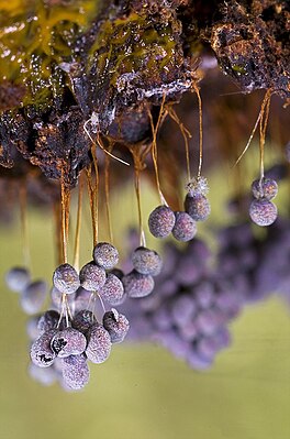
Drooping fruiting bodies of Badhamia utricularis |
||||||||||
| Systematics | ||||||||||
|
||||||||||
| Scientific name | ||||||||||
| Myxogastria | ||||||||||
| LSOlive |
The Myxogastria , usually referred to as Myxomycetes , sometimes also referred to as real slime molds or plasmodial slime molds , are a subgroup of the Amoebozoa . They include around 900 species in almost 60 genera .
All species go through several morphologically very different phases during their life, from microscopic cells to slimy, amorphous organisms that are visible to the naked eye and sometimes unusually conspicuously shaped fruit bodies. In doing so, they can reach considerable sizes and weights.
The group is distributed worldwide, but it is much more common in temperate latitudes and has a higher species richness than in polar regions, the subtropics or the tropics. They are mainly found in light forests, but can also be found in extreme locations such as deserts, under closed snow cover or even under water.
Features and life cycle


The haplo-diplontic life cycle of the Myxogastria can roughly be divided into three phases, first a unicellular , mononuclear , haploid phase, which ends with the formation of diploid zygotes. Then follows a stage as amorphous, multinucleated, diploid plasmodium and finally the reproductive phase with the formation of the diploid fruiting body , the so-called fructification, in which haploid spores are endogenously formed again. At each stage, the respective appearance changes drastically.
Single-cell, single-core phase
Spurs
The spores are haploid , mostly round and measure between 5 and 20, rarely up to 24 micrometers in diameter. Their surface is usually network-like, burr, warty to prickly structured, only very rarely is it smooth. The structure also creates the perceived coloration of the spore mass, the spores are not pigmented . In some species (especially in the Badhamia genus ) the spores form clumps. The "color" of the spores, their shape, the sculpting and the diameter are important features for determination.
The most important factors for germination are humidity and temperature; it is unclear what other factors play a role. The spores usually remain viable for several years, individual spores of herbarium material germinated even after 75 years. After their formation, the spores initially contain a diploid cell nucleus, and meiosis takes place in the spore. During germination, the spore coats either open along special germ pores or crevices or tear irregularly and then release one to four haploid protoplasts .
Myxamoeba and myxoflagellates
In species that reproduce sexually, haploid cells germinate from the spores . A myxamoeba or a myxoflagellate germinates from the spore, depending on the ambient conditions: Myxamoeba move amoeboid , i.e. crawl on the substrate and arise in rather dry conditions. Myxoflagellates are flagellated and buoyant, they arise in rather damp to wet surroundings. Myxoflagellates almost always have two flagella, one flagella is usually shorter and in some cases can be almost completely reduced. The flagella are not only used for locomotion, but are also used to whip up food. If the humidity regime changes, the cells can switch between the two manifestations. Both forms do not have a cell wall. Like the next, this stage of development is used for nutrition and is therefore referred to as the first trophic stage (nutritional stage). In this phase, Myxogastria feed on bacteria and fungal spores and probably also solutes. In this unicellular stage, they multiply through simple cell division .
If the environmental conditions change adversely during this phase (for example extreme temperatures, severe drought or lack of food), Myxogastria can switch to an extremely long-lasting, thin- shelled state of persistence, the so-called microcyst . To do this, the myxamoeba round off and secrete a thin-walled cell wall. In this condition, they can easily survive for a year or more. If the living conditions improve, they become active again.
Zygote formation
When two cells of the same type meet at this stage, they fertilize each other by fusing protoplasms and cell nuclei to form a diploid zygote . The conditions that trigger this are not known. From the diploid zygote, through multiple nuclear division without subsequent cell division, a multinucleated plasmodium emerges. If the output cells were still flagellated, they change their shape from the flagellated form to the myxamoeba before merging. For the formation of a zygote, cells of different crossing types are necessary ( heterothallia ).
Plasmodium
The second trophic stage begins with the formation of the plasmodium . The polynuclear organism now absorbs as many nutrients as possible by phagocytosis . These are bacteria, protists , solutes, molds and higher fungi as well as small particles of organic material. They enable the cell to grow enormously and to divide the cell nucleus many times over, so that the cell is soon visible to the naked eye and - depending on the type - can occupy an area of up to one square meter (an artificially grown cell from Physarum polycephalum achieved Area of 5.5 square meters). In their trophic stage as Plasmodium, Myxogastria species always have numerous cell nuclei: in the small, non-veined protoplasmodia there are 8 to 100 nuclei, in the large veined networks 100 to 10 million nuclei. However, all of them are still part of a single cell, which has a viscous, slimy consistency and is either transparent, white or brightly colored (orange, yellow, pink).
In this phase, the cell has chemotactic and (negative) phototactic abilities, so it is mobile and moves on substances such as nutrients to or from dangerous substances and light sources. The movement is caused by the granular plasma that pulsates in one direction within the cell. The cell achieves an unusually high speed of up to 1000 micrometers (1 mm) per second (for comparison: in plant cells this speed is 2 to 78 micrometers per second). At this stage, too, a persistence stage can be formed, the so-called sclerotium , a hardened, resistant form made up of numerous so-called macrocysts that affect the myxogastria at this stage, for example. B. enable survival of winters or drought.
Fructification


Mature plasmodia can then develop fruiting bodies under appropriate circumstances. The exact reasons that trigger this process are not known; laboratory observations suggest changes in humidity, temperature or pH value as well as periods of hunger as triggering factors for some species. The plasmodium stops eating and crawls - now attracted by the light (= "positive phototaxis") - to a dry, bright spot, from where the later spread of the spores can optimally take place. Once the fructification has started, it cannot be reversed. If there are disturbances, spore-bearing fruit bodies are formed, but these are often malformed.
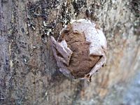
The fruiting bodies emerging from the Plasmodium or parts of it can be smaller than a millimeter, but in extreme cases they can also reach sizes of up to one meter and a total weight of up to 20 kilograms ( Brefeldia maxima ). Mostly they have the shape of pedunculated or sessile sporangia (whereby the peduncles are not cellular), but can also appear as veined to reticulated plasmodiocarps , pillow-shaped ethers or apparently pillow-shaped pseudo - ethers . The fruiting bodies almost always have a hypothallus at the base. The masses of spores produced in the fruiting bodies are found in almost all species (exceptions are the Liceida species and some representatives of the Echinostelium genus ) in a reticulate to thread-like structure, the so-called capillitium .
After the opened fruiting bodies have dried off, the spores are mainly spread by the wind; small animals such as woodlice , mites or beetles also play a certain role, which either emerge from contact with fruiting bodies with spores or take them up as part of their food and excrete them again capable of germination. Spreading through rivers plays only a minor role .
Asexual forms
Some Myxogastria have asexual forms that reproduce apomictically . These are all diploid. No meiosis takes place before the spores germinate and the formation of plasmodium takes place without any fusion of two cells.
Distribution and ecology
distribution
Myxogastria are widespread worldwide, evidence exists from all continents. In principle, however, detailed statements about the distribution of individual taxa of the Myxogastria suffer from the fact that large parts of the world have only been insufficiently or not yet investigated. Only Europe and North America are considered to be at least fundamentally developed floristically. The results so far suggest that a cosmopolitan distribution can be assumed for the majority of all species.
They occur most frequently and with the greatest biodiversity in temperate latitudes, much less frequently in polar regions or the subtropics and tropics. The physical properties of the substrate as well as climatic conditions are decisive for the presence of the species in certain locations; endemism is rare.
The distribution area extends in the north to Alaska , Iceland , northern Scandinavia , Greenland and northern Russia . These are not just individual, special species; an overview study was able to identify more than 150 different species for the arctic and subarctic regions of Iceland, Greenland, Northern Russia and Alaska alone. The tree line is clearly exceeded, in Greenland the distribution area even reaches the 77th parallel.
In the forests of temperate zones, the Myxogastria then reach their greatest biodiversity and their highest abundance, where the ideal conditions are found in the form of the abundant presence of organic material, the open forests with sufficient but not too high humidity and for nivicole (= snow-dwelling) species many areas with a long period of closed snow cover.
Only a few species colonize the tropics and subtropics. The main reasons given for the lower frequency in tropical forests are consistently high levels of humidity, which on the one hand prevent the necessary drying out of the fruiting bodies (disadvantageous for the spread of the spores) and on the other hand promote an infestation by mold . Other factors are the relative lack of light (impairment of positive phototaxis), calm, acidic soils, a variety of predators and frequent heavy rains that wash away or destroy the cells. Above all, the extensive groups of soil and deadwood-dwelling species decline with increasing moisture. In a comparative study of Costa Rica , the findings provide the relatively dry Tropical Moist Forest 73% of the overall findings, those from the already significantly wetter Tropical Premontane Wet Forests only 18% and in the rainforest type Lower Premontane Rainforest he dropped to only 9 % Proportion of.
In the Southern Arctic Circle, there are only isolated records from the Southern Shetland Islands , the Southern Orkney Islands , South Georgia and the Antarctic Peninsula . The extremely small number of Antarctic or sub-Antarctic records compared to the Arctic Circle illustrates how unexplored the myxomycete flora of this area is. Up to 1983 only five finds were known, even after that there were only a few other finds. However, the only two thorough studies of myxomycete flora in the region indicate significantly higher numbers of species in general. Samples from sub-Antarctic forests of the Argentine Patagonia and Tierra del Fuego were able to detect 67 species, a survey on the Macquarie Island, which is only 128 square kilometers in size, already 22.
Habitats

The majority of all known Myxogastria species live terrestrially in open forests. The most important microhabitat is dead wood , and the bark of living trees, rotting plant material from the litter horizon, soil and animal excrement are also important.
In contrast to this, Myxogastria can be found in numerous unusual places. The extensive group of so-called nivicolored Myxogastria grows under closed snow cover in order to quickly fructify and release their spores when exposed (for example during a thaw). Deserts ( 33 species have been recorded for the Sonoran Desert alone) or living leaves of plants in the tropics are also habitats of Myxogastria.
The aquatic way of life of some species is unusual. Individual representatives of the genera Didymium , Physarum , Perichaena , Fuligo , Comatricha and Licea could be observed living under water both as myxoflagellates and as plasmodia. They only came to fructification when the water receded or left it, with the exception of the species Didymium difforme , for which evidence of submerged fructification was provided.
Relationships with other living beings
The relationships between the Myxogastria and other living beings have only been explored to a limited extent. As predators, a number of are arthropods known especially beetles (like the families of Rove , sponge ball beetle , rhysodidae , eucinetidae , point beetle , rail bugs , dust fungus beetles , smooth bark beetles , Moder beetle ), mites and springtails . Various nematodes have also proven to be predators; they anchor themselves with their rear end in the plasma strands of plasmodia or even live within the strands.
The Diptera also some specialists have emerged, mostly representatives of fungus gnats and the fungus gnats and fruit flies . The species Epicypta testata is particularly common , it is found in many cases on Enteridium lycoperdon , Enteridium splendens , Lycogala epidendrum and Tubifera ferruginosa .
Some real mushrooms have specialized in the colonization of Myxogastria, almost always they are representatives of the hose fungus . Verticillium rexianum is most frequently found (mostly on species of Comatricha or Stemonitis ), while on species of Physarida one can often find Gliocladium album or Sesquicillium microsporum . Polycephalomyces tomentosus is often found among representatives of the Trichiida . Nectriopsis violacea specializes in yellow tan blossom ( Fuligo septica ) .
In some investigations, bacteria mostly from the Enterobacteriaceae family were found living on Plasmodium as so-called “bacterial associates” . Only the combination of plasmodia and bacterial associates is it possible to bind atmospheric nitrogen or to produce enzymes that enable the decomposition of lignin , carboxymethyl celluloses or xylan , for example . In individual cases, the plasmodia acquired salt or heavy metal tolerances through the association .
Taxonomy
nomenclature
The name Myxogastria (= "slime bellies") was introduced in 1970 by Lindsay Shepherd Olive , referring to the Myxogastridae described as a family by Thomas Huston Macbride as early as 1899 . The name appears similarly in 1829 with Elias Magnus Fries , who called all slime molds "Myxogasteres".
Due to competing systematics and the parallel use of the mutually exclusive botanical and zoological regulations, there are many different names with similar or even identical cuts (for example Myxogastromycetidae, Mycetozoa, Myxomycota). The best known is certainly the name "Myxomycetes", first described by Johann Heinrich Friedrich Link in 1833 , which, although no longer recognized as a taxon today, became a trivial name as "Myxomycetes" and is still used in scientific publications today. The common name "Schleimpilze", which misleadingly suggests a relationship to the fungi, is incorrectly applied to this group, since it denotes the higher taxon of the Eumycetozoa (which is no longer recognized today) .
scope
The group includes around 900 to 1000 species. A survey in 2000, aimed at completeness, resulted in 1012 validly described taxa , including 866 at species level. Another study in 2007 estimated the number of species to be well over 1000, after which the Myxogastria, as by far the largest group of slime molds, comprised well over 900 species. The continuous description of new taxa makes it clear that knowledge of the group is still incomplete. Estimates based on sequenced environmental samples assume that the group with a total of 1200–1500 species is significantly larger than previously known. Of the known 1012 taxa, only a few species are common: 305 are known from a single location or only a single collection, 258 species were collected between 2 and 20 times in a few locations and only 446 of the species were common with more than 20 found in several locations .
Numerous new descriptions suffer from the fact that Myxogastria can be morphologically influenced by environmental influences and at the same time have only a few diagnostic features with little variety of forms. In combination with the "problematic habit" of authors to willingly describe a new taxon based on only a few available specimens, this leads to numerous duplicates (occasionally even at the genus level as in the case of Squamuloderma nullifila , actually a species of the genus Didymium ).
Systematics and phylogeny
With regard to the Myxogastria itself, the following system is based on Adl et al. 2005, however, in the ranks and the further subdivision, it takes up the systematics of Dykstra and Keller 2000, in which the Myxogastria are addressed as "Mycetozoa".
The sister taxon are the dictyostelia . Together with them and the Protostelia , the Myxogastria formed the taxon of the slime mold (Eumycetozoa) according to the classical view. The myxogastria can be separated from the other two groups mainly through the formation of the fruiting bodies: while in Protostelia a separate fruiting body is created from each individual and always mononuclear cell, Dictyostelia formed cell associations consisting of individual cells, so-called pseudoplasmodia, which then form convert to fruit bodies. According to more recent understanding, the superordinate taxon of the Eumycetozoa can no longer be maintained.
- Order Liceida
- Liceidae family
- Family Listerelliidae
- Family Enteridiidae
- Order Echinosteliida
- Echinosteliedae family
- Family Clastodermidae
- Order Trichiida
- Family Dianemidae
- Family Trichiidae
- Order Stemonitida
- Family Stemonitidae
|
Myxogastria cladogram
|
- Order Physarida
- Family Elaeomyxidae
- Family Physaridae
- Family Didymiidae
It largely corresponds to the traditional classification that has been in use since the work of Lister and Lister at the beginning of the 20th century. Molecular genetic studies confirm this classification and consolidate it with reliable results. The most basic group are the Echinosteliida, the other groups can also be summarized in two super clusters, which can also be morphologically delimited according to the color of the spores.
Fossil record
Fossil finds that are assigned to the Myxogastria are extremely rare. Due to their short-lived nature and the sensitive structure of the plasmodia and fruiting bodies, Myxogastria are out of the question for fossilization or similar processes; only their spores can mineralize . The few known fossils of the life stages are therefore all only known as inclusions in amber .
Up to 2010 fruit bodies had been described three times, two spores and one Plasmodium. Two older taxa ( Charles Eugène Bertrand's Myxomycetes mangini and Bretonia hardingheni from 1892) are considered dubious and are usually no longer taken into account today.
In 1952, Friedrich Walter Domke described a 35 to 40 million year old find made of Baltic amber of the species Stemonitis splendens, which still occurs today . The state of preservation and the completeness of the fruiting bodies, which allow an almost perfect determination, is remarkable. From the same period and also using Baltic amber, Heinrich Dörfelt and Alexander Schmidt first described Arcyria sulcata in 2003 , a species that is very similar to today's Arcyria denudata . Both finds indicate that the fruiting bodies of the Myxogastria have changed little over the past 35 to 40 million years.
In contrast, the Protophysarum balticum , described in 2006 by Dörfelt and Schmidt from Baltic amber , is considered questionable. Not only that it contradicted the description of the genre in many points and, due to the lack of a Latin diagnosis, is not a valid publication . Its character as a type of Myxogastria was quickly questioned because - in contrast to previous finds - important details of the fruiting bodies were not visible or contradicted the classification. Therefore, today it is more likely that the find is a lichen from the area of the genus Chaenotheca . The only previously reported find of a Plasmodium comes from Dominican amber and was assigned to the Physarida . However, it is also considered dubious, especially since the publication is classified as insufficient due to deficiencies.
The only known mineralized fossils are two spore finds known in 1971, one of which was classified as being related to the post-glacial period as Trichia favoginea . The suggestion to include Myxogastria spores in pollen and spore analyzes , however, remained without echo.
Research history

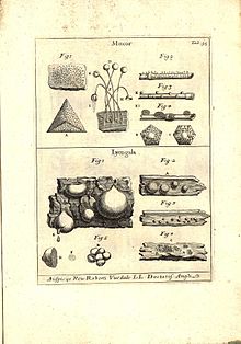
Due to their inconspicuous nature, the Myxogastria have only been specifically explored late. Thomas Panckow first mentioned a myxogastria, namely the blood milk fungus , under the name "Fungus cito crescentes", in 1654 in his Herbarium Portatile, or agile book of herbs and plants accompanied by an illustration, so it understands it as a mushroom. In 1729 Pier Antonio Micheli first formulated the idea of the previously known species as a group of living beings that can be distinguished from mushrooms, which Heinrich Friedrich Link helped establish from 1833. Elias Magnus Fries had already documented the plasmodial stage in 1829, and Anton de Bary was then able to observe the stage of spore germination in 1864. Since he was also able to observe the plasma flow in the cell that enabled movement, he saw them as closer to the animals and consequently described them anew as Mycetozoa, literally translated as "mushroom animals", a view that dominated until the second half of the 20th century.
From 1874 to 1876 Jósef Tomasz Rostafinski , a student of De Bary, published the first comprehensive monograph on the group. In 1894, 1911 and 1925, Arthur and Guilielma Lister published three monographs that are still received today as “exemplary, fundamental works”, as does The Myxomycetes by Thomas H. Macbride and George Willard Martin , published in 1934 . Important works of the late 20th century were the monographs by George Willard Martin and Constantine John Alexopoulos in 1969 and Lindsay S. Olive in 1975. The former in particular is considered to be important because it “began the modern era of the taxonomy of Myxogastria”. Persoon, Rostafinski, Lister, Macbride as well as Martin and Alexopoulos contributed in particular to the knowledge of new taxa and their systematics; their work significantly increased the number of species.
In addition to the monographs dealing with the group worldwide, some large-scale flora are also of importance, especially since the group has significantly less regionally effective restrictions due to its disjoint distribution. Important large florets are z. B. Robert Hagelsteins The Mycetozoa of North America (1944) and Marie Farrs Band for Flora Neotropica 1973, more recent works of this kind are Bruce Ings The Myxomycetes of Britain and Ireland , Lado and Pandos Band for Flora Mycologica Iberica from 1997 and Yamamoto's The Myxomycete Biota of Japan from 1998. The creation of the three-volume work Die Myxomyceten Deutschlands and the neighboring Alpine region with special emphasis on Austria , which was written by Hermann Neubert , Wolfgang Nowotny and Karlheinz Baumann from 1993 to 2000 and published by himself , is unusual . Although the authors were formally amateurs, their work received very positive reviews from scholars and is now a much-cited standard work. In 2011 the work Les Myxomycètes was published , consisting of a text and an illustrated book. The authors are Michel Poulain , Marianne Meyer and Jean Bozonnet . This work, too, achieved the status of a standard work shortly after its publication.
Myxogastria and human
The Myxogastria are largely of no economic or cultural significance for humans. Physarum polycephalum is a common model organism in biology for studying cell motility, cell growth and cell differentiation. The ease of cultivation and the size of the cell are advantageous for handling.
Yellow tufted plasmodia ( Fuligo septica ) and Enteridium lycoperdon ethers are eaten in the Veracruz area of Mexico. There they are grilled under the name “caca de luna” , in English “moon poop”, known as a delicacy.
proof
- ^ HO Schwantes: Biology of mushrooms . Stuttgart 1996, ISBN 3-8252-1871-6 , pp. 308-311
- ↑ a b c d e f g h i j Document for the paragraph: Wolfgang Nowotny: Myxomyceten (Schleimpilze) and Mycetozoa (mushroom animals) - life forms between animals and plants In: Wolfgang Nowotny (Ed.): Wolfsblut und Lohblüte. Life forms between animals and plants = Myxomycetes (= Stapfia . Band 73 ). Linz 2000, ISBN 3-85474-056-5 , p. 7–37 (German, English, French, Spanish).
- ↑ a b c d e Sina M. Adl, Alastair GB Simpson, Mark A. Farmer, Robert A. Andersen, O. Roger Anderson, John A. Barta, Samuel S. Bowser, Guy Brugerolle, Robert A. Fensome, Suzanne Fredericq , Timothy Y. James, Sergei Karpov, Paul Kugrens, John Krug, Christopher E. Lane, Louise A. Lewis, Jean Lodge, Denis H. Lynn, David G. Mann, Richard M. McCourt, Leonel Mendoza, Øjvind Moestrup, Sharon E. Mozley-Standridge, Thomas A. Nerad, Carol A. Shearer, Alexey V. Smirnov, Frederick W. Spiegel, Max FJR Taylor: The New Higher Level Classification of Eukaryotes with Emphasis on the Taxonomy of Protists. The Journal of Eukaryotic Microbiology 52 (5), 2005; S. 399-451 ( The New Higher Level Classification of Eukaryotes with Emphasis on the Taxonomy of Protists. Accessed on 28 December 2019 . )
- ^ H. Raven, Ray F. Evert and Helena Curtis: Biology of Plants. 2nd Edition. Walter de Gruyter GmbH, 1988, ISBN 3-11-011476-3 , p. 267-269 .
- ↑ a b c d e f Reference for the paragraph: Henry Stempen, Steven L. Stevenson: Myxomycetes. A Handbook of Slime Molds . Timber Press, 1994, ISBN 0-88192-439-3 , pp. 15-18 .
- ↑ a b c d e Wolfgang Nowotny: Myxomycetes (Schleimpilze) and Mycetozoa (mushroom animals) - life forms between animals and plants In: Wolfgang Nowotny (Ed.): Wolfsblut und Lohblüte. Life forms between animals and plants = Myxomycetes (= Stapfia . Band 73 ). Linz 2000, ISBN 3-85474-056-5 , p. 7–37 (German, English, French, Spanish).
- ↑ Wolfgang Richter: Old Schleimer . In: ZEIT Wissen, 01/2007, online
- ↑ a b Proof of the paragraph: Sina M. Adl, Alastair GB Simpson, Mark A. Farmer, Robert A. Andersen, O. Roger Anderson, John A. Barta, Samuel S. Bowser, Guy Brugerolle, Robert A. Fensome, Suzanne Fredericq, Timothy Y. James, Sergei Karpov, Paul Kugrens, John Krug, Christopher E. Lane, Louise A. Lewis, Jean Lodge, Denis H. Lynn, David G. Mann, Richard M. McCourt, Leonel Mendoza, Øjvind Moestrup , Sharon E. Mozley-Standridge, Thomas A. Nerad, Carol A. Shearer, Alexey V. Smirnov, Frederick W. Spiegel, Max FJR Taylor: The New Higher Level Classification of Eukaryotes with Emphasis on the Taxonomy of Protists. The Journal of Eukaryotic Microbiology 52 (5), 2005; S. 399-451 ( The New Higher Level Classification of Eukaryotes with Emphasis on the Taxonomy of Protists. Retrieved on December 28, 2019 . )
- ↑ a b c d Henry Stempen, Steven L. Stevenson: Myxomycetes. A Handbook of Slime Molds . Timber Press, 1994, ISBN 0-88192-439-3 , pp. 49-58 .
- ↑ Steven L. Stephenson, Martin Schnittler, Carlos Lado: Ecological characterization of a tropical myxomycete assemblage — Maquipucuna Cloud Forest Reserve, Ecuador In: Mycologia, Vol. 96, pp. 488-497, 2004
- ↑ Proof of the paragraph: Steven L. Stephenson, Yuri K. Novozhilov, Martin Schnittler: Distribution and Ecology of Myxomycetes in High-Latitude Regions of the Northern Hemisphere In: Journal of Biogeography, 27: 3, 2000, pp. 741-754
- ↑ Reference for the paragraph: Steven L. Stephenson, Martin Schnittler, Carlos Lado: Ecological characterization of a tropical myxomycete assemblage — Maquipucuna Cloud Forest Reserve, Ecuador In: Mycologia, Vol. 96, pp. 488-497, 2004
- ^ A b J. Putzke, AB Pereira, MTL Putzke: New Record of Myxomycetes to the Antarctica. In: V Simposio Argentino y I Latinoamericano de Investigaciones Antarticas, 2004, Buenos Aires - Argentina, Actas del V Simposio Argentino y I Latinoamericano de Investigaciones Antarticas , Vol. 1, pp. 1-4, 2004, PDF online ( Memento from 14. October 2012 in the Internet Archive )
- ↑ a b B. Ing, RIL Smith: Further myxomycete records from South Georgia and the Antarctic peninsula. In: British Antarctic Survey Bulletin 59, pp. 80-81, 1983
- ↑ Diana W. de Basanta, Carlos Lado, Arturo Estrada-Torres, Steven L. Stephenson: Biodiversity of myxomycetes in subantarctic forests of Patagonia and Tierra del Fuego, Argentina In: Nova Hedwigia, 90: 1-2, pp. 45-79
- ↑ SL Stephenson, GA Laursen, RD Seppelt: Myxomycetes of subantarctic Macquarie Island. In: Australian Journal of Botany 55, pp. 439-449
- ↑ a b Reference for the paragraph: Henry Stempen, Steven L. Stevenson: Myxomycetes. A Handbook of Slime Molds . Timber Press, 1994, ISBN 0-88192-439-3 , pp. 49-58 .
- ↑ Uno H. Eliasson: Myxomycetes on living leaves in the tropical rainforest of Ecuador; an investigation based on the herbal material of higher plants In: Wolfgang Nowotny (Hrsg.): Wolfsblut und Lohblüte. Life forms between animals and plants = Myxomycetes (= Stapfia . Band 73 ). Linz 2000, ISBN 3-85474-056-5 , p. 81 (German, English, French, Spanish).
- ↑ Documentation for the paragraph: Lora A. Lindley, Steven L. Stephenson, Frederick W. Spiegel: Protostelids and myxomycetes isolated from aquatic habitats In: Mycologia, Vol. 99, pp. 504–509, 2007
- ^ Rolf G. Beutel, Richard AB Leschen: Handbuch der Zoologie - Coleoptera, Beetles, Volume 1: Morphology and Systematics (Archostemata, Adephaga, Myxophaga, Polyphaga partim) . 1st edition. de Gruyter , 2005, ISBN 3-11-017130-9 , p. 304 (English).
- ^ Rolf G. Beutel, Richard AB Leschen: Handbuch der Zoologie - Coleoptera, Beetles, Volume 1: Morphology and Systematics (Archostemata, Adephaga, Myxophaga, Polyphaga partim) . 1st edition. de Gruyter , 2005, ISBN 3-11-017130-9 , p. 269 (English).
- ^ Rolf G. Beutel, Richard AB Leschen: Handbuch der Zoologie - Coleoptera, Beetles, Volume 1: Morphology and Systematics (Archostemata, Adephaga, Myxophaga, Polyphaga partim) . 1st edition. de Gruyter , 2005, ISBN 3-11-017130-9 , p. 147 (English).
- ^ Rolf G. Beutel, Richard AB Leschen: Handbuch der Zoologie - Coleoptera, Beetles, Volume 1: Morphology and Systematics (Archostemata, Adephaga, Myxophaga, Polyphaga partim) . 1st edition. de Gruyter , 2005, ISBN 3-11-017130-9 , p. 433 (English).
- ^ Rolf G. Beutel, Richard AB Leschen: Handbuch der Zoologie - Coleoptera, Beetles, Volume 1: Morphology and Systematics (Archostemata, Adephaga, Myxophaga, Polyphaga partim) . 1st edition. de Gruyter , 2005, ISBN 3-11-017130-9 , p. 438 (English).
- ^ Richard AB Leschen, Rolf G. Beutel, John F. Lawrence: Handbuch der Zoologie - Coleoptera, Beetles, Volume 2: Morphology and Systematics (Elateroidea, Bostrichiformia, Cucujiformia partim) . de Gruyter, 2010, ISBN 978-3-11-019075-5 , p. 61 (English).
- ^ Richard AB Leschen, Rolf G. Beutel, John F. Lawrence: Handbuch der Zoologie - Coleoptera, Beetles, Volume 2: Morphology and Systematics (Elateroidea, Bostrichiformia, Cucujiformia partim) . de Gruyter, 2010, ISBN 978-3-11-019075-5 , p. 300 (English).
- ^ Richard AB Leschen, Rolf G. Beutel, John F. Lawrence: Handbuch der Zoologie - Coleoptera, Beetles, Volume 2: Morphology and Systematics (Elateroidea, Bostrichiformia, Cucujiformia partim) . de Gruyter, 2010, ISBN 978-3-11-019075-5 , p. 424 (English).
- ^ Richard AB Leschen, Rolf G. Beutel, John F. Lawrence: Handbuch der Zoologie - Coleoptera, Beetles, Volume 2: Morphology and Systematics (Elateroidea, Bostrichiformia, Cucujiformia partim) . de Gruyter, 2010, ISBN 978-3-11-019075-5 , p. 483 (English).
- ^ Courtney M. Kilgore, Harold W. Keller: Interactions Between Myxomycete Plasmodia and Nematodes , In: Inoculum 59: 1, pp. 1-3, 2008
- ↑ a b c Reference for the paragraph: Henry Stempen, Steven L. Stevenson: Myxomycetes. A Handbook of Slime Molds . Timber Press, 1994, ISBN 0-88192-439-3 , pp. 59-68 .
- ↑ Reference for the paragraph: Indira Kalyanasundaram: A Positive Ecological Role for Tropical Myxomycetes in Association with Bacteria In: Systematics and Geography of Plants, 74: 2, pp. 239–242, 2004
- ^ Lindsay S. Olive: The Mycetozoa: A Revised Classification , In: Botanical Review, 36: 1, pp. 59-89, 1970
- ^ Hermann Neubert , Wolfgang Nowotny , Karlheinz Baumann : The Myxomycetes of Germany and the neighboring Alpine region with special consideration of Austria . tape 1 . Karlheinz Baumann Verlag, Gomaringen 1993, ISBN 3-929822-00-8 , p. 11 .
- ↑ a b c d M. Schnittler & DW Mitchell: Species Diversity in Myxomycetes Based on the Morphological Species Concept - a Critical Examination In: Wolfgang Nowotny (Ed.): Wolfsblut und Lohblüte. Life forms between animals and plants = Myxomycetes (= Stapfia . Band 73 ). Linz 2000, ISBN 3-85474-056-5 , p. 39–53 (German, English, French, Spanish).
- ↑ Proof of the paragraph: Sina M. Adl, Brian S. Leander, Alastair GB Simpson, John M. Archibald, O. Roger Anderson, David Bass, Samuel S. Bowser, Guy Brugerolle, Mark A. Farmer, Sergey Karpov, Martin Kolisko, Christopher E. Lane, Deborah J. Lodge, David G. Mann, Ralf Meisterfeld, Leonel Mendoza, Øjvind Moestrup, Sharon E. Mozley-Standridge, Alexey V. Smirnov, Frederick Spiegel: Diversity, Nomenclature, and Taxonomy of Protists . Systematic Biology, Vol. 56, 2007, pp. 684-689.
- ↑ a b Jim Clark: The species problem in the myxomycetes In: Wolfgang Nowotny (Ed.): Wolfsblut und Lohblüte. Life forms between animals and plants = Myxomycetes (= Stapfia . Band 73 ). Linz 2000, ISBN 3-85474-056-5 , p. 39–53 (German, English, French, Spanish).
- ↑ Michael J. Dykstra, Harold W. Keller: Mycetozoa In: John J. Lee, GF Leedale, P. Bradbury (Eds.): An Illustrated Guide to the Protozoa . tape 2 . Allen, Lawrence 2000, ISBN 1-891276-23-9 , pp. 952-981 .
- ↑ Anna Maria Fiore-Donno, Sergey I. Nikolaev, Michaela Nelson, Jan Pawlowski, Thomas Cavalier-Smith, Sandra L. Baldauf: Deep Phylogeny and Evolution of Slime Molds (Mycetozoa) In: Protist, 161: 1, p. 55– 70, 2010
- ↑ Adl, SM, Simpson, AGB, Lane, CE, Lukeš, J., Bass, D., Bowser, SS, Brown, MW, Burki, F., Dunthorn, M., Hampl, V., Heiss, A. , Hoppenrath, M., Lara, E., le Gall, L., Lynn, DH, McManus, H., Mitchell, EAD, Mozley-Stanridge, SE, Parfrey, LW, Pawlowski, J., Rueckert, S., Shadwick, L., Schoch, CL, Smirnov, A. and Spiegel, FW: The Revised Classification of Eukaryotes. Journal of Eukaryotic Microbiology , 59: 429-514, 2012, PDF Online
- ↑ A.-M. Fiore-Donno, C. Berney, J. Pawlowski, SL Baldauf: Higher-Order Phylogeny of Plasmodial Slime Molds (Myxogastria) Based on Elongation Factor 1-A and Small Subunit rRNA Gene Sequences. In: Journal of Eukaryotic Microbiology, 52, pp. 201-210, 2005
- ↑ Documentation for the paragraph: A.-M. Fiore-Donno, C. Berney, J. Pawlowski, SL Baldauf: Higher-Order Phylogeny of Plasmodial Slime Molds (Myxogastria) Based on Elongation Factor 1-A and Small Subunit rRNA Gene Sequences. In: Journal of Eukaryotic Microbiology, 52, pp. 201-210, 2005
- ↑ a b c d Harold W. Keller, Sydney E. Everhart: Myxomycete species concepts, monotypic genera, the fossil record, and additional examples of good taxonomic practice In: Revista Mexicana de Micología, 27, 2008, pp. 9-19
- ↑ a b Reference to the paragraph: Alan Graham: The role of myxomyceta spores in palynology (with a brief note on the morphology of certain algal zygospores) In: Review of Palaeobotany and Palynology, 11: 2, pp. 89-99, 1971
- ^ BM Waggoner, GO Poinar: A Fossil Myxomycete Plasmodium from Eocene-Oligocene Amber of the Dominican Republic. In: Journal of Eukaryotic Microbiology, 39, pp. 639-642, 1992
- ^ Reference for the paragraph: Henry Stempen, Steven L. Stevenson: Myxomycetes. A Handbook of Slime Molds . Timber Press, 1994, ISBN 0-88192-439-3 , pp. 19-21 .
further reading
- Hermann Neubert, Wolfgang Nowotny, Karlheinz Baumann, Heidi Marx: The Myxomycetes of Germany and the neighboring Alpine region with special consideration of Austria. Vol. 1–3, Karlheinz Baumann Verlag, Gomaringen.
- George W. Martin, Constantine John Alexopoulos, Marie Leonore Farr: The genera of Myxomycetes , Iowa City, 1983, ISBN 0-87745-124-9 .
- Wolfgang Nowotny (Ed.): Wolfsblut and Lohblüte. Life forms between animals and plants = Myxomycetes (= Stapfia . Band 73 ). Linz 2000, ISBN 3-85474-056-5 (German, English, French, Spanish). online on ZOBODAT
- Henry Stempen, Steven L. Stevenson: Myxomycetes. A Handbook of Slime Molds . Timber Press, 1994, ISBN 0-88192-439-3 .


