Big liver fluke
| Big liver fluke | ||||||||||||
|---|---|---|---|---|---|---|---|---|---|---|---|---|

Fasciola hepatica |
||||||||||||
| Systematics | ||||||||||||
|
||||||||||||
| Scientific name | ||||||||||||
| Fasciola hepatica | ||||||||||||
| Linnaeus , 1758 |
The large liver fluke ( Fasciola hepatica ) is a parasite ( sucking worm ) that occurs worldwide, up to 3 cm in length and a bay leaf-like shape, which, as a final host, attacks herbivores such as cattle or sheep , but also pigs , humans and other mammals (it was also found in the Dog feces detected). Infection in sheep occurs mainly in summer and autumn on pasture. The infestation with the great liver fluke is called fasciolosis .
Development cycle
An adult liver fluke lays eggs in the bile duct system of the definitive host, which are released into the environment with the faeces . These eggs survive there for 2 to 6 months. They are 130–150 µm long, 62–90 µm wide and have a characteristic yellowish color with an operculum , but cannot be distinguished from eggs of the species Fasciola gigantica . The eggs ripen in the water. If the climatic conditions are favorable (15-25 ° C), the eyelash larvae ( miracidia ) develop within 9-21 days . No further development takes place below 10 ° C, but the eggs remain viable for months.
After hatching, the eyelash larvae swim with the help of their cilia until they find an intermediate host. This must be done within 24 hours, otherwise the miracidia will die. The amphibious dwarf mud snail ( Lymnaea truncatula , also known as the “liver fluke snail ”, <1 cm in length) acts as an intermediate host . In Australia the intermediate host is Lymnaea tomentosa , in North America it is Fossaria modicella and Fossaria bulimoides . Gerhard Piekarski mentions Galba and Radix as further genera ( see lung snails ).
In the snail, the larva develops into a sporocyst , in which several redia are formed, which in turn form daughter redia until these tail larvae ( cercaria ) have developed. This process takes about 2 months and ends in up to 2000 cercariae. The cercariae develop in the snail best at temperatures of 20 to 25 ° C within 6 to 7 weeks.
The cercaria actively leave the snail and attach themselves to plants or similar just below the surface of the water, where they encyst. During this process, they lose their tail. The metacercarials are infectious 24 hours after encysting . The survival time at +10 ° C is at least 130 days; at −10 ° C for 7–28 days; at −20 ° C for 12 h, and at +35 ° C for up to 14 days. Survival even under European winter conditions appears possible. Direct sunlight and high humidity at extreme temperatures seem to affect the survival time.
From these plants they in turn reach the ultimate host and can grow there. Rare infections in humans in Europe are known or suspected through consumption of raw watercress , more rarely also dandelions and windfalls , especially from contaminated pastureland . Within an hour of receiving the metacercariae start in the small intestine to excystation of the host. After penetrating the wall of the small intestine, they can be found in the abdominal cavity within 2 hours. After 24 hours, the majority develop into maturing worms ( larvae ), after 48 hours these begin to penetrate the liver capsule , the majority have reached the liver parenchyma within 6 days. In the liver they migrate for 5 to 6 weeks, feeding directly on liver tissue. They eventually migrate into the bile ducts, where they reach sexual maturity. The oviposition begins (according to examinations in sheep and cattle) approx. 2 months after ingestion of the metacercariae. 2–3 weeks for the eggs to mature, 6 to 7 weeks for the cercariae to develop in the snails, results in a cycle duration of 14 to 23 weeks.
distribution
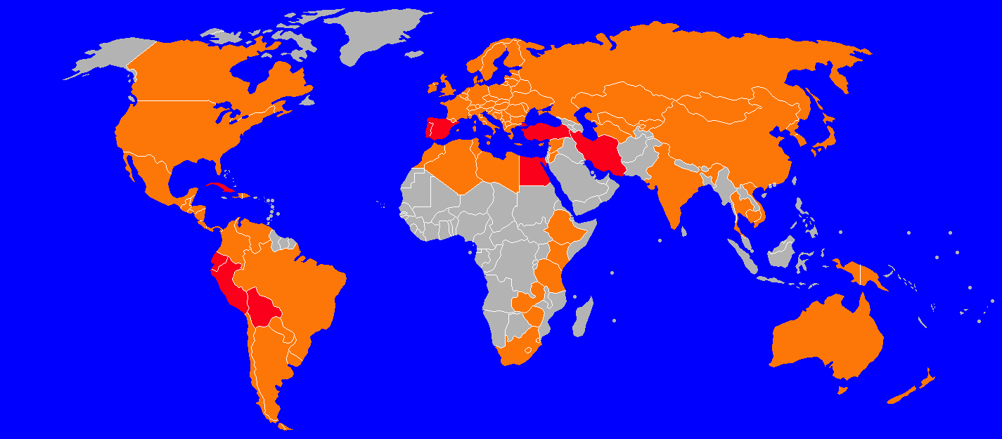
The world map shows the distribution of the liver fluke. Countries colored red have a high prevalence. In Europe these are Spain and Portugal, in Asia Turkey and Iran, in Africa Egypt, in Latin America Cuba, Bolivia, Peru and Equador. The highlands of Bolivia have the highest prevalence in the world. Orange colored indicate a medium or low prevalence. The graphic shows the state of knowledge from 2011.
Web links
- with overview graphic of the development cycle
- Extensive text: The Life cycle of Fasciola hepatica, Stewart J. Andrews ( Memento from March 28, 2006 in the Internet Archive ) (PDF, 205 kB)
- G. Dreyfuss, P. Vignoles, M. Abrous, D. Rondelaud: Unusual snail species involved in the transmission of Fasciola hepatica in watercress beds in central France. In: Parasite. Volume 9, Number 2, June 2002, pp. 113-120, ISSN 1252-607X . PMID 12116856 .
- When humans consume contaminated plants or water ...
- Fasciola ( CDC )
Individual evidence
- ^ S. Mas-Coma: Epidemiology of fascioliasis in human endemic areas . In: Journal of Helminthology . tape 79 , no. 3 , September 2005, ISSN 0022-149X , p. 207-216 , doi : 10.1079 / JOH2005296 ( cambridge.org [accessed May 16, 2020]).
- ^ Robert W Tolan: Fascioliasis Due to Fasciola hepatica and Fasciola gigantica Infection: An Update on This 'Neglected' Neglected Tropical Disease . In: Laboratory Medicine . tape 42 , no. 2 , February 2011, ISSN 0007-5027 , p. 107–116 , doi : 10.1309 / LMLFBB8PW4SA0YJI ( oup.com [accessed May 16, 2020]).
- ↑ DP McMANUS, JP Dalton: Vaccines against the zoonotic trematodes Schistosoma japonicum, Fasciola hepatica and Fasciola gigantica . In: Parasitology . tape 133 , S2, October 2006, ISSN 0031-1820 , p. S43 – S61 , doi : 10.1017 / S0031182006001806 ( cambridge.org [accessed May 16, 2020]).
