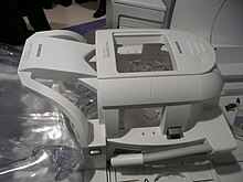
Functional magnetic resonance imaging
The functional magnetic resonance tomography , abbreviated fMRT or fMRI (for English functional magnetic resonance imaging ), is an imaging procedure to display physiological functions inside the body with the methods of magnetic resonance tomography (MRT). fMRI in the narrower sense refers to processes which can display activated brain areas (mostly based on blood oxygenation) with high spatial resolution; In the broader sense, other functional imaging techniques such as dynamic cardiac MRT, time-resolved MRT examination of joint movements or perfusion MRT are also referred to as functional MRT. Sometimes the procedure or its result is also referred to as a brain scan .
introduction

With fMRI recordings, it is possible to visualize changes in blood flow in areas of the brain that are attributed to metabolic processes that are in turn related to neuronal activity. The different magnetic properties of oxygenated and deoxygenated blood are used here ( BOLD contrast ). The activation of cortical areas leads to an increase in metabolism, as a result of which the activated area reacts with a disproportionate increase in blood flow (so-called neurovascular coupling ). This increases the concentration of oxygenated (diamagnetic) relative to deoxygenated (paramagnetic) hemoglobin . Via the intermolecular electron dipole-nuclear dipole relaxation mechanism , this change in concentration causes a change in the effective transverse relaxation time of the observed hydrogen nuclear spins and thus leads to a signal change in the MRT . In order to draw conclusions about the location of a neural activity, the magnetic resonance signal of the tissue is compared at two points in time - e.g. B. in the stimulated or experimental state on the one hand and in the rest or control state on the other hand. The recordings can be compared with one another using statistical test methods and the statistically significant differences (which correspond to the stimulated areas) can be spatially assigned and displayed.
An fMRI examination usually consists of three phases:
- Prescan: a short, low-resolution scan. This can be used to check the correct positioning of the patient.
- Anatomical MRT scan: a spatially high-resolution scan in order to be able to show the anatomy of the area to be examined in detail via image fusion .
- The actual fMRI scan: a quick scan that uses the BOLD contrast to show differences in blood flow in the tissue being examined.
When examining the brain for experimental purposes, the subject can be presented with a repeated stimulus in the third partial scan, for example. The stimulus is often linked to a task for the test person, such as the request to press a button for each object X shown . What most of the experiments have in common is the frequent repetition of the task. Statistical methods can then be used to compare recorded data from the stimulus phase with those from the resting phase. The difference calculated from this is then projected in false colors onto the previously performed anatomical MR scan.
Neurology and neuropsychology in particular benefit from the possibilities of fMRI. For example, through comparative studies with fMRI between people who suffer from mental disorders such as depression , anxiety and obsessive-compulsive disorders, and healthy control persons, clear and z. Sometimes chronic differences in brain metabolism can be demonstrated.
historical development
Linus Pauling had already discovered in 1935 that the magnetic properties of hemoglobin change depending on the degree of oxygenation. This effect forms the basis for measuring brain activity with functional MRI, which was developed in the 1980s and 1990s. In 1982 Keith Thulborn and co-workers showed that hemoglobin in blood samples appears differently in its MRI signal depending on the degree of oxygenation. The same observation was 1990 by Seiji Ogawa and co-workers in vivo made in Sprague-Dawley rats; the property of hemoglobin to cause different MRI signals was called the "blood oxygenation level dependent (BOLD)" effect. First human fMRI results showing brain activity after visual stimulation were published in 1991 by John W. Belliveau and co-workers.
Limits
Compared to the other established non-invasive neurophysiological examination methods, such as EEG , the (relatively young) fMRI shows significantly more powerful possibilities in spatial-localizing examination, but a fundamentally much lower temporal resolution. An additional uncertainty arises from the indirect character of the method - the neural activity is not measured directly, but inferred from changes in blood flow and oxygenation. A roughly linear relationship between stimuli that are longer than four seconds and the BOLD effect is assumed. Whether the BOLD effect reliably reproduces neuronal activity with shorter stimuli is debatable and is still the subject of current research.
Further technical limitations of the fMRI measurement are:
- In intact tissues, the BOLD effect is not only caused by the blood in the vessels, but also by the cell tissue around the vessels.
- If the measurement voxel falls below a minimum size when measuring the BOLD effect , vessels that have a cross-section that is larger than the specified voxel size can be incorrectly interpreted as neuronal activity.
In addition, there is criticism of the basic assumptions and possible findings from fMRI studies, based on the fact that the visualization of the measurement data of the fMRI has a constructive component, which means that the researchers' model ideas can be represented rather than actual processes. Furthermore, statistical correction calculations to exclude random results were missing in numerous investigations.
According to a study published in 2016, many scientists did not adequately check the requirements for using the statistical software to be used. This leads to false positive signals and shows activity in the brain where there is none. Many of the more recent studies (several thousand could be affected) that looked at thought processes and emotions and combined measurements from several test subjects could be worthless.
See also
- Neuroethics
- AFNI , FSL - software packages for processing and evaluation
literature
- Scott A. Huettel, Allen W. Song, Gregory McCarthy: Functional Magnetic Resonance Imaging . 2nd Edition. Palgrave Macmillan, 2008, ISBN 978-0-87893-286-3 .
- NK Logothetis , J. Pauls, M. Augath, T. Trinath, A. Oeltermann: Neurophysiological investigation of the basis of the fMRI signal. In: Nature. 412, 2001, pp. 150-157.
- Robert L. Savoy: Functional MRI , in Encyclopedia of the Brain, Ramachandran (Ed). Academic Press (2002).
Individual evidence
- ↑ Frank Schneider, Gereon R. Fink (ed.): Functional MRT in psychiatry and neurology . Springer, Berlin 2007, ISBN 3-540-20474-1 ( limited preview in the Google book search).
- ↑ Michael Graf, Christian Grill, Hans-Dieter Wedig (eds.): Acceleration injury of the cervical spine: cervical whiplash . 1st edition. Steinkopff, Berlin 2008, ISBN 978-3-7985-1837-7 , p. 160–161 ( limited preview in Google Book search).
- ↑ Gabriele Benz-Bohm (Ed.): Pediatric Radiology . 2nd Edition. Thieme, Stuttgart 2005, ISBN 3-13-107492-2 , p. 239 ( limited preview in Google Book search).
- ↑ Alexandra Jorzig; Frank Sarangi: Digitization in Healthcare: A Compact Foray through Law, Technology and Ethics . Springer Berlin Heidelberg, May 22, 2020, ISBN 978-3-662-58306-7 , p. 114–.
- ↑ Claudia Steinbrink; Thomas Lachmann: Reading and Spelling Disorders: Basics, Diagnostics, Intervention . Springer-Verlag, April 22, 2014, ISBN 978-3-642-41842-6 , p. 39–.
- ^ L. Pauling: The oxygen equilibrium of hemoglobin and its structural interpretation . In: Proc Natl Acad Sci USA . tape 21 , no. 4 , 1935, pp. 186-191 , PMID 16587956 .
- ^ KR Thulborn, JC Waterton, PM Matthews, GK Radda: Oxygenation dependence of the transverse relaxation time of water protons in whole blood at high field . In: Biochim Biophys Acta . tape 714 , no. 2 , 1982, p. 265-270 , doi : 10.1016 / 0304-4165 (82) 90333-6 , PMID 6275909 .
- ^ S. Ogawa , TM Lee, AR Kay, DW Tank : Brain magnetic resonance imaging with contrast dependent on blood oxygenation . In: Proc Natl Acad Sci USA . tape 87 , no. 24 , 1990, pp. 9868-9872 , PMID 21247060 .
- ↑ JW Belliveau, DN Kennedy, RC McKinstry, BR Buchbinder, RM Weisskoff, MS Cohen, JM Vevea, TJ Brady, BR Rosen: Functional mapping of the human visual cortex by magnetic resonance imaging . In: Science . tape 254 , 1991, pp. 716-719 , doi : 10.1126 / science.1948051 , PMID 1948051 .
- ↑ Yevgeniy B. Sirotin, Aniruddha Das: Anticipatory haemodynamic signals in sensory cortex not predicted by local neuronal activity . In: Nature . tape 457 , p. 475-479 , doi : 10.1038 / nature07664 , PMID 19158795 .
- ↑ AM Dale, RL Buckner: Selective averaging of rapidly presented individual trials using fMRI . In: Human Brain Mapping . tape 5 , no. 5 , 1997, pp. 329-340 , doi : 10.1002 / (SICI) 1097-0193 (1997) 5: 5 <329 :: AID-HBM1> 3.0.CO; 2-5 , PMID 20408237 .
- ^ S. Ogawa, TM Lee, AS Nayak, P. Glynn: Oxygenation-sensitive contrast in magnetic resonance image of rodent brain at high magnetic fields . In: Magn Reson Med . tape 14 , no. 1 , 1990, p. 68-78 , doi : 10.1002 / mrm.1910140108 , PMID 2161986 .
- ↑ J. Frahm, KD Merboldt, W. Hänicke: Functional MRI of human brain activation at high spatial resolution . In: Magn Reson Med . tape 29 , no. 1 , 1993, p. 139-144 , doi : 10.1002 / mrm.1910290126 , PMID 8419736 .
- ↑ Veronika Hackenbroch: cerebral voodoo. In: Der Spiegel . 18/2011, May 2, 2011.
- ↑ Craig M. Bennett, Abigail A. Baird, Michael B. Miller, George L. Wolford: Neural Correlates of Interspecies Perspective Taking in the Post-Mortem Atlantic Salmon: An Argument For Proper Multiple Comparisons Correction. In: JSUR. 1 (1), 2010, pp. 1-5. (PDF; 864 kB) ( Memento of the original from March 4, 2016 in the Internet Archive ) Info: The archive link was inserted automatically and has not yet been checked. Please check the original and archive link according to the instructions and then remove this notice.
- ^ Hanno Charisius : Illusions in the brain scan. In: Süddeutsche Zeitung . July 6, 2016, p. 16.
- ↑ Anders Eklund, Thomas E. Nichols, Hans Knutsson: Cluster failure: Why fMRI inferences for spatial extent have inflated false-positive rates . In: Proceedings of the National Academy of Sciences . tape 113 , no. 28 , June 28, 2016, ISSN 0027-8424 , p. 7900-7905 , doi : 10.1073 / pnas.1602413113 ( pnas.org [accessed April 24, 2019]).
Web links
- Easy to understand introduction to the topic of fMRI
- fmri for newbies English-language, extensive site on fMRI
- Article Functional Magnetic Resonance Imaging on Scholarpedia (English)
- Cerebral Voodoo Article by Veronika Hackenbroch, Der Spiegel 18/2011 from May 2, 2011

