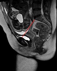
Birth mechanics
As birth mechanics refers to the mechanical interaction between the fetus and the birth canal during birth .
Anatomical basics
Birth canal anatomy
The birth canal , i.e. the part through which the child has to pass during birth in order to be born, is formed on the one hand by the maternal pelvis and on the other hand by maternal soft tissues ( cervix , vagina , vulva ). While the soft tissue is stretchable to a certain extent, the bony pelvis is relatively firm. Although the influence of hormones during pregnancy leads to a loosening of the symphysis (= pubic symphysis ) in order to widen the pelvis a little, this mechanism is limited. The bony pelvic structures are therefore decisive for the mechanics of childbirth on the maternal side.
The small pelvis is anatomically divided into four classic levels:
- The pelvic entrance has a transversely oval shape (it is wider than it is deep) and is formed by the so-called promontory of the sacrum , the pubic symphysis and the iliac bone . The shortest distance between the posterior symphysis surface and the promontory, which is known as the conjugata vera obstetrica , is decisive for the mechanics of the birth .
- The pelvic width is formed by the posterior wall of the symphysis and the middle of the third sacral vertebra. It is round and the largest level of the birth canal.
- The pelvic narrowing is formed at the front by the lower edge of the symphysis, at the back by the transition from the sacrum to the coccyx (articulatio sacrococcygis) and laterally by the spinous processes of the ischial bone (spinae ischiadicae). This narrow is longitudinally oval, so it is deeper than it is wide.
- The pelvic outlet is finally formed by Symphysenunterrand, the ischial tuberosities and the tip of coccyx.
In obstetrics , however, for the sake of simplicity, Hodge's parallel planes are used, which are each 4 cm apart and are based on the spinous processes of the ischial bone (spinae ischiadicae) as a reference or zero plane.
Anatomy of the fetus
The child's head as the part of the fetus that has the greatest circumference is decisive in terms of birth mechanics. Furthermore, the head is longer than it is wide - the distance from the forehead to the back of the head is longer than the distance between the two temples - i.e. longitudinally oval. In contrast to the mother's pelvis, the child is not a rigid system, but the head can be rotated, bent or stretched relatively freely from the trunk. Furthermore, the child's skull bones are not yet firmly ossified and thus the head can be compressed to a certain extent. After the head, the child's shoulder and pelvis are the widest parts.
Course of birth
The passage of the child through the birth canal is a passive process from a child's point of view and adapts to the respective shape and width of the birth canal according to the anatomical conditions. The head follows the law of least compulsion . From a mechanical point of view, the birth proceeds as follows:
- Entry of the head into the small pelvis . Since the pelvic entrance runs transversely oval, the head is also positioned transversely. The child's face therefore points to the right or to the left.
- Flexion . The head moves deeper into the pelvic cavity and passively flexes it. The so-called small fontanel , which lies on the back of the child's head, comes to the front (in the "leading line")
- 1. rotation . The head now gets into the pelvic narrowing and has to rotate 90 ° according to its shape in order to move the occiput forward. This process is supported by the maternal levator muscle . The face now points backwards and the head now comes to the pelvic outlet.
- Elongation . The head must now pass through a kink in the birth canal. To do this, he braces himself against the edge of the symphysis and thus reaches a stretched position. The occiput appears under the symphysis. In the so-called incision , the vertex, forehead and head pass through the perineum .
- 2. Rotation . After the head is born, the shoulders follow. Since these are wider than deep, in contrast to the head, the child has to turn 90 ° again, the child's face at the end points in the same direction as it was when entering the pelvis.
- Birth of the trunk . This process takes place without tension. For this purpose, the child is developed following the curved birth canal around the symphysis on the mother's stomach.
Adjustment anomalies of the child's head can complicate this normal birth process.
literature
- Klaus Diedrich u. a. (Ed.): Gynecology and Obstetrics . Springer-Verlag Berlin, Heidelberg, New York 2000, ISBN 3-540-65258-2 .
