Dermatome (anatomy)
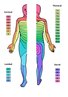
The dermatome (from ancient Greek δέρμα dérma ' skin ' and ancient Greek τομή tomḗ '(cut)') is the segmental area of skin that is sensitively innervated by a spinal cord nerve ( spinal nerve ) .
Embryological basics
During the segmentation of the embryo in the trunk area from the paraxial mesoderm , to the side of the neural tube and the chorda dorsalis , initially the primary vertebra ( thus ) arise . The dermis is formed from its dorsolateral part . This plant is also known as a dermatome . As a result of this segmental origin of the individual skin areas, there is also a segmental assignment to the corresponding spinal nerve.
anatomy
This segmental assignment of the spinal cord nerves to the corresponding skin areas is also retained in adults. The cell bodies of these sensitive neurons lie outside the spinal cord in the spinal ganglion ( ganglion spinal ). However, there is an overlap of the skin areas of the individual spinal nerves. If a spinal nerve is damaged, there is therefore no complete loss of sensitivity in the relevant dermatome. The overlap should be less pronounced for the perception of pain and temperature stimuli than of touch stimuli. Due to the overlap, a noticeable loss of sensitivity often only occurs when two adjacent segments fail.
Autonomous area
In the area of the neck and in the lumbar and cruciform area, the ventral rami of the spinal nerves form plexuses :
- Cervical plexus ( plexus cervicalis )
- Arm plexus ( brachial plexus )
- Lumbar and cross braid ( plexus lumbosacralis )
The fiber exchange in these braids creates s. G. Plexus nerves. In most cases, these lead to nerve cell processes in several segments. An area of skin that is only sensitively innervated by a certain nerve is also called the autonomous area of this nerve. Damage to the nerve typically leads to a loss of sensitivity in its autonomous area.
Selected autonomous areas:
- Ulnar nerve : little finger
- Median nerve : terminal phalanx of the index and middle fingers
- Radial nerve : above the metacarpal bone of the thumb (but not constant)
- Nervus fibularis profundus : first space between the toes
- Tibial nerve : sole of the foot and heel
Transferred pain
Since viscerosensitive ( viscera "intestines", "inner organs") sensations are transmitted via the spinal nerves, but these cannot be assigned to a precise location due to lack of experience in the responsible region of the cerebral cortex , these pains from the cerebrum are (incorrectly) corresponding sensitive skin areas , usually assigned to that of the same spinal nerve, for example shoulder pain in epigastric peritonitis . In diseases of internal organs, hypersensitivity to external stimuli can arise in a certain area of the skin, the so-called Head's zone . This phenomenon is examined in diagnostics as a head zone sample (also Kalchschmidt sample). There may also be transferred pain in the muscles ( myotome ) of the corresponding segment ( Mackenzie zone ). An example of this is referred pain in angina pectoris .
The Head's zone (also Head zone), named after the English neurologist Sir Henry Head (1861-1940) is defined as the area of skin in the indented due to the body structure (→ metamerism ) a current via the associated spinal cord segment cross-connection between the somatic and the autonomic nervous system . Certain internal organs are assigned to this area, see the following table. Head's zone, which is assigned to a specific organ, can extend over several dermatomes, but has a reflexively significant maximum point. Irritation of the associated internal organ can result in a mostly equilateral pain zone via a viscerocutaneous reflex (hyperalgesia zone). This phenomenon is called referred pain . The pain can u. U. to spread to neighboring segments or the entire body half (generalization). Some alternative medical methods are supposed to use a reversal of the reflex process to influence internal organs by influencing certain skin zones mechanically, thermally or pharmacologically. These methods, for which there is no scientific evidence, are called reflex therapies .
| organs | Dermatome | Body side |
|---|---|---|
| heart | C 3-4, Th 1-5 | predominantly left, also right arm |
| Thoracic aorta | C 3-4, Th 1-7 | both sides |
| Pleura | Th 2-12 | the respective half of the body (ipsilateral) |
| Lungs | C 3-4 | ipsilateral |
| esophagus | Th 1-8 | both sides |
| stomach | Th (5) 6-9 | Left |
| Liver and biliary tract | Th (5) 6-9 (10) | right |
| pancreas | Th 6-9 | forward Left |
| Duodenum | Th 6-10 | right |
| Jejunum | Th 8-11 | Left |
| Ileum | Th 9-11 | both sides |
| Cecum , proximal colon | Th 9-10, L 1 | right |
| distal colon | Th 9 - L 4 | Left |
| rectum | Th 9 - L 4 | Left |
| Kidney and ureter | Th 9 - L 1 (2) | ipsilateral |
| Adnexa | Th 12 - L 4 | ipsilateral |
| peritoneum | Th 5-12 | both sides |
| spleen | Th 6-10 | Left |
See also
Individual evidence
- ↑ R. Putz, R. Pabst (Ed.): Sobotta, Atlas of the Anatomy of Man. Volume 2, 20th edition. Urban & Schwarzenberg, Munich 1993, ISBN 3-541-17370-X , p. 346.
- ↑ E Ernst, P Posadzki, MS Lee: Reflexology: an update of a systematic review of randomized clinical trials . In: Maturitas . 68, No. 2, February 2011, pp. 116-120. doi : 10.1016 / j.maturitas.2010.10.011 .
- ↑ Norbert Boss (Ed.): Roche Lexicon Medicine. 2nd Edition. Hoffmann-La Roche AG and Urban & Schwarzenberg, Munich 1987, ISBN 3-541-13191-8 , p. 744.

