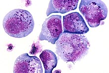
Inclusion bodies

Inclusion bodies or aggregates of protein ( English inclusion bodies ) are small, after staining under the light microscope visible particles in the interior of cells . They consist of collections of mostly incorrectly or incompletely folded proteins , which are produced, among other things, in excessive synthesis and precipitate in the cell nucleus or cytoplasm .
properties
Whereas overexpression of recombinant proteins spherical inclusion bodies often have formed in E. coli .mu.m a diameter of 0.2 to 1.5, and accumulate up to a proportion of over 50% of total cell protein, which may even lead to a deformation of the cell. As a rule, there is only one inclusion body per bacterial cell, whereby this is never membrane-bound but has a porous structure. For a long time it was assumed that inclusion bodies were an unstructured collection of polypeptides, but the latest research suggests a defined conformation even in the aggregated form. Likewise, it has long been assumed that the aggregates are insoluble in vivo , but it is now generally accepted that the aggregates can also dissolve in vivo . Refolding by chaperones into the native form has even been demonstrated. The inclusion bodies usually consist of fully synthesized proteins which, however, are only partially folded. The proportion of active protein in the aggregates can be very high despite the non- native form. In the case of β-glucosidase, up to 30% and in the case of endoglucanase almost 100% of the protein present in the inclusion body can be present in an active conformation.
Occurrence
Inclusion bodies are often used as a diagnostic criterion in viral infections. Their synthesis can also be stimulated in a targeted manner and the resulting protein can be further processed industrially, for example in genetically modified organisms , i.e. the production of recombinant proteins.
In mammals , inclusion bodies are found in protein misfolding diseases , e.g. B. in nerve cells in the form of Pick bodies in Pick's disease , beta-amyloid plaques in Alzheimer's disease , Huntingtin in Huntington 's disease , in prion diseases and in various hereditary diseases ( Lafora disease , Fabry disease ). Inclusion bodies also occur in various bacterial ( chlamydia , Escherichia coli ) and viral infectious diseases ( viroplasm in polyoma , smallpox , adeno , asfar and herpes viruses , yellow fever , rabies , distemper , panleucopenia , Sendai virus infection ). In pharmacology, therefore, the suitability of chemical and pharmacological chaperones as therapeutic approaches is also being investigated.
In birds , inclusion bodies are also found in polyoma ( polyomavirus infection of parrots ) and herpes virus infections ( infectious laryngotracheitis ).
Inclusion bodies can also be detected in plant viruses (e.g. Lily Mottle virus , Potyviridae ).
Individual evidence
- ↑ F Baneyx, M Mujacic: Recombinant protein folding and misfolding in Escherichia coli . In: Nature biotechnology , vol. 22, no. 11, 2004, pp. 1399-1408, PMID 15529165
- ^ S Ventura, A Villaverde: Protein quality in bacterial inclusion bodies . In: Trends in biotechnology , 2006, vol. 24, no. 4, pp. 179-185, PMID 16503059
- ↑ JL Corchero, R Cubarsí, S Enfors, A. Villaverde: Limited in vivo proteolysis of aggregated proteins . In: Biochemical and Biophysical Research Communications , 1997, vol. 237, issue 2, pp. 325-330, PMID 9268709
- ↑ H Lethanh, P Neubauer, F Hoffmann: The small heat-shock proteins IbpA and IbpB reduce the stress load of recombinant Escherichia coli and delay degradation of inclusion bodies . In: Microbial cell factories , 2005, vol. 4, no. 1, p. 6, PMID 15707488
