Polyomaviridae
| Polyomaviridae | ||||||||||||||
|---|---|---|---|---|---|---|---|---|---|---|---|---|---|---|

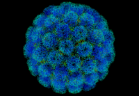
3D model of the SV40 capsid |
||||||||||||||
| Systematics | ||||||||||||||
|
||||||||||||||
| Taxonomic characteristics | ||||||||||||||
|
||||||||||||||
| Left | ||||||||||||||
|
The Polyomaviridae family comprises non-enveloped DNA viruses that cause persistent infections in various vertebrates ( mammals , rodents, and birds ) and in humans . These and the closely related family Papillomaviridae were created by splitting up the former family Papovaviridae and were elevated to the rank of a class as Papovaviricetes by the International Committee on Taxonomy of Viruses (ICTV) in March 2020 in order to combine the two families of the papovaviruses. The originally only genus Polyomavirus was already divided into four genera Alphapolyomavirus to Deltapolyomavirus ( see below ). The family name is derived from the Greek πολύς ( poly : many, several) and the suffix -oma from the name for tumors , as the first virus of the family to be identified, the murine polyomavirus , leads to various tumors in newborn mice.
In 2013, hybrids between papillomaviruses and polyomaviruses were described.
morphology
The virions (virus particles) of the polyomaviruses consist of a bare capsid , about 40 to 45 nm in diameter , which is composed of 72 capsomeres . The capsomeres are arranged in an icosahedral symmetry ( T = 7 ). The individual capsomeres are formed at the base from five molecules of the capsid protein VP1 ( pentamer ), which, however, are not arranged uniformly but twisted ( skewed ). One speaks therefore of a twisted, icosahedral symmetry (T = 7d). On the inside of the capsid, the capsid proteins VP2 and VP3 stabilize the VP1 framework; they also interact with the dsDNA inside the capsid. Different virus particles are often observed, including empty, normally structured capsids, very small, empty capsids (microcapsids) and irregular tubular structures that are formed from the capsid proteins in different compositions. The VP1 capsid protein can spontaneously assemble without further virus proteins to form virus-like particles which, however, cannot pack nucleic acids. In the real Virion, the VP1 makes up around 70% of the total protein content.
Inside the capsids is the covalently closed DNA ring of the virus genome. As with the Papillomaviridae, this is twisted several times ( “supercoiled” ) and, together with cellular histones, forms a nucleoprotein complex that is structurally very similar to the eukaryotic nucleosomes . Of the five known histones, the histones H2a , H2b , H3 and H4 are found .
The capsids are very environmentally stable and can not be inactivated with diethyl ether , 2-propanol or detergents (soap) . They are heat-stable up to 50 ° C for 1 hour; in the simultaneous presence of magnesium chloride in 1 M concentration, the capsids are unstable, which, as with the papilloma viruses, indicates the dependence of the capsid structure on divalent cations .
Genome
The polyomavirus genome consists of a single molecule of a double-stranded, covalently closed ring of DNA. Starting from a non-coding, regulatory region, the open reading frames (ORFs) for the 5 to 9 different virus proteins are arranged in such a way that the ORFs for the early transcripts run in one reading direction and those for the late transcripts in the opposite direction. The early transcripts include the large and possibly also small T antigen and other regulatory proteins; the late transcripts code for the three structural proteins VP1-3. The reading frames overlap with partially different reading frames, so that the polyomaviruses with a genome size of approximately 4.7 to 5.5 kbp code for a relatively large number of proteins. Like the papilloma viruses, the polyomaviruses do not have their own DNA polymerase to replicate the viral DNA; they also rely on the cell's own polymerases. The viral early proteins bind to the enhancers and promoters for their own reading frames in the regulatory region. This suppresses the production of these proteins in favor of the later proteins in the course of virus replication. The investigation of this mechanism in SV-40 led to the discovery of these regulatory sequences in eukaryotes and to the development of the enhancer concept in molecular biology.
Biological importance
Avian polyomaviruses, for example, trigger the French moult . The BK virus (BKV, BK polyomavirus or BKPyV) can lead to the loss of the transplant in humans with immunosuppressive treatment after kidney transplantation . The BK virus can also cause respiratory infection or cystitis in children, hemorrhagic cystitis in bone marrow transplant recipients, ureteral stenosis in kidney transplant recipients, and meningoencephalitis in AIDS patients.
The BK and JC viruses belonging to this genus, which are also referred to as human polyomavirus 1 and 2 (formerly Polyomavirus hominis type 1 and 2), persist in kidney tissue; in the normal population, 100% of antibodies against BK virus (BKV) and about 80% against JC virus (JCV) can be detected.
The fact that an infection with these viruses only extremely rarely leads to death in people who are not significantly pre-damaged and if there is no double infection or secondary infection shows, on the one hand, that these disease-causing viruses are very well adapted to humans as their reservoir hosts. Damage to its reservoir host is not an advantageous effect for a virus, since it depends on it for its own reproduction. The diseases caused by this virus in the reservoir host are ultimately only side effects of the infection. Secondly, it also makes it clear that humans have also been able to adapt to this virus over the course of many generations. At the moment there is a BK virus infection of 80-90% of the population. The JC virus leads to progressive multifocal leukoencephalopathy (PML) in cellular immunosuppressed patients ( AIDS St. C3 ). PML is almost always fatal.
The Simiane Virus 40 or SV40 is a potential cause of various tumor diseases. Parts of the SV40 DNA are used in molecular biology as a particularly powerful promoter or enhancer .
Systematics

The former genus Polyomavirus was split into four parts, which are preceded by the names of the Greek letters alpha to delta ( ICTV as of November 2018):
- Family Polyomaviridae
-
- Genus Alphapolyomavirus
- Species mouse polyomavirus
( Mus musculus polyomavirus 1 , Murines polyomavirus , old Parotid tumor virus , MPyV) - Species Chlorocebus pygerythrus polyomavirus 1
( green monkey polyomavirus 1 , vervet monkey polyomavirus 1 , AGMPyV-1) - Species Chlorocebus pygerythrus polyomavirus 3
( green monkey polyomavirus 3 , vervet monkey polyomavirus 3 , AGMPyV-3) - Species Papio cynocephalus polyomavirus 1
( baboon polyomavirus 1 , BPyV-1) - Species Mesocricetus auratus polyomavirus 1
( hamster polyomavirus , HaPyV) - Species Pan troglodytes polyomavirus 1 to 7
( chimpanzee polyomavirus 1 to 7 ) - Species Mus musculus polyomavirus 1 ( Murine Polyomavirus , MPyV)
- Species mouse polyomavirus
- Genus Betapolyomavirus (temporarily separated into the genera Orthopolyomavirus and Wukipolyomavirus )
- Species Human Polyomavirus 1
( Polyomavirus hominis 1 , BK virus , en. BK polyomavirus , officially Human polyomavirus 1 , HPyV-1, BKPyV or BKV) - Species Human Polyomavirus 2
( Polyomavirus hominis 2 , JC virus , en. JC polyomavirus , officially Human polyomavirus 2 , HPyV-2, JCPyV or JCV) - Species Human Polyomavirus 3
( Polyomavirus hominis 3 , KI virus , en.KI polyomavirus , officially Human polyomavirus 3 , HPyV-3, KIPyV or KIV) - Species Human Polyomavirus 4
( Polyomavirus hominis 4 , WU virus , en. WU Polyomavirus , officially Human polyomavirus 4 , HPyV-4, WUPyV or WUV) - Species human polyomavirus 5
( Merkel cell polyomavirus , MCPyV) - Species Macaca mulatta polyomavirus 1
( Simian virus 40 , MmPV1 or SV40) - Species Chlorocebus pygerythrus polyomavirus 2
( green monkey polyomavirus 2 , vervet monkey polyomavirus 2 , AGMPyV-2) - Species Papio cynocephalus polyomavirus 2
( baboon polyomavirus 2 , BPyV-2)
- Species Human Polyomavirus 1
- Genus Gammapolyomavirus (obsolete Avipolyomavirus )
- Species Aves polyomavirus 1
( avian polyomavirus 1 , budgerigar polyomavirus , budgerigar fledgling disease virus , or BFPyV BFDV) - polyomavirus the nestling of the disease Wellensittiche - Species Corvus monedula polyomavirus 1
( crow polyomavirus ) - Species Anser anser polyomavirus 1 ( haemorrhagic polyomavirus of geese , GHPV)
- Species Serinus canaria polyomavirus 1
( finch polyomavirus )
- Species Aves polyomavirus 1
- Genus Delta Polyomavirus
- Species Human polyomavirus 6 ( Human polyomavirus 6 , HPyV-6)
- Species Human polyomavirus 7 ( Human polyomavirus 7 , HPyV-7)
- Species Human polyomavirus 10 ( Human polyomavirus 10 , HPyV-10)
- Species Human polyomavirus 11 ( Human polyomavirus 11 , HPyV-11)
- Not assigned to any genus within the Polyomaviridae
- Species Bos taurus polyomavirus 1
( Bovine Polyomavirus , BPyV) - Species rabbit polyomavirus
( Rabbit kidney vacuolating virus , RKV) - Species Murine Pneumotropic Virus (MPtV)
- Species Baboon polyomavirus 1
( Simian virus 12 , SV12)
- Species Bos taurus polyomavirus 1
literature
- J. Hou et al. : Family Polyomaviridae . In: CM Fauquet, MA Mayo et al. : Eighth Report of the International Committee on Taxonomy of Viruses . London / San Diego 2005, ISBN 0-12-249951-4 , pp. 231-238
- MJ Imperiale, EO Major: Polyomaviridae . In: David M. Knipe, Peter M. Howley (eds.-in-chief): Fields' Virology . Volume 2. 5th edition. Philadelphia 2007, ISBN 0-7817-6060-7 , pp. 2263-2298
Web links
- Polyomaviridae (NCBI)
Individual evidence
- ↑ a b c d e f ICTV: ICTV Taxonomy history: Human polyomavirus 1 , EC 51, Berlin, Germany, July 2019; Email ratification March 2020 (MSL # 35)
- ↑ Annabel Rector, Marc Van Ranst: Animal papillomaviruses , Virology Volume 445, Issue 1–2, October 2013, pp. 213-223, doi: 10.1016 / j.virol.2013.05.007
- ↑ José Carlos Mann Prado, Telma Alves Monezi, Aline Teixeira Amorim, Vanesca Lino, Andressa Paladino, Enrique Boccardo: Human polyomaviruses and cancer: an overview . In: Clinics (Sao Paulo), 73 (Suppl 1), p. E558s, doi: 10.6061 / clinics / 2018 / e558s , PMC 6157077 (free full text), PMID 30328951 (online September 26, 2018), Fig. 1
- ↑ Ugo Moens, Sébastien Calvignac-Spencer, Chris Lauber, Torbjörn Ramqvist, Mariet CW Feltkamp, Matthew D. Daugherty, Ernst J. Verschoor, Bernhard Ehlers: ICTV Virus Taxonomy Profile: Polyomaviridae . In: J Gen Virol. , 98 (6), June 22, 2017, pp. 1159–1160, doi: 10.1099 / jgv.0.000839 , PMC 5656788 (free full text), PMID 28640744
- ↑ dsDNA Viruses> Polyomaviridae . In: ICTV Report, June 2017, revised in July 2018, Table 2A
- ↑ SIB: Alphapolyomavirus , on: ViralZone
- ↑ SIB: Betapolyomavirus , on: ViralZone
- ↑ SIB: Gammapolyomavirus , on: ViralZone
- ↑ SIB: Deltapolyomavirus , on: ViralZone
