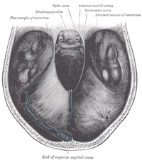
Tentorium cerebelli
The tentorium cerebelli (dt. Cerebellar tent ) is a horizontal, coarse meningeal structure inside the skull, which divides its interior into a supratentorial (above the tentorium) and an infratentorial (below the tentorium) space. Immediately above this is the occipital lobe of the cerebrum , and below it is the cerebellum .
anatomy
The tentorium cerebelli is a connective tissue formation of the inner layer ( lamina interna ) of the hard meninges ( dura mater ). It covers the posterior cranial fossa ( fossa cranii posterior ) like a roof and is located in the fissura transversa cerebri (formerly also known as fissura telencephalicodiencephalis ).
The cerebellar tent extends laterally between the temporal bone and the transverse sinus . It is fixed dorsally on the internal occipital protuberance , laterally on both sides at the edges of the transverse sinus sulcus and rostrally on the superior margin of the temporal bone pyramid and runs near the apex of the temporal bone over the trigeminal impression , from there it runs medially to the dorsum sellae , where it is reaches the processus clinoideus posterior and further rostrally with a branch the processus clinoideus anterior . It forms the so-called plicae petroclinoideae anteriores et posteriores on these structures . At the sulcus sinus transversus the root of the tentorium encompasses the sinus transversus and at the upper edge of the temporal bone the sinus petrosus superior . Medially, between the legs of the tentorium, the structure known as the incisura tentorii (tentorium slit ) is formed, through which the mesencephalon with the arteriae cerebri posteriores , nervi trochleares and nervi oculomotorii passes. The bottom of the Tentoriumschlitzes forms the dorsum sellae The notch tentorii is rostrally from the sphenoid bone is limited and forms the only connection between the supratentorial (above the tentorium located) and the infratentorial (below the tentorium located) subarachnoid space in which cerebrospinal fluid is .
The transition between falx cerebri and tentorium cerebelli, where the rectus sinus is located, is located on the midline of the tent ridge . Together with the falx cerebri, the cerebellar tent forms a tension belt system that mechanically stabilizes the skull capsule from the inside. The tentorium cerebelli carries the dorsal half of the telencephalon and thus prevents its pressure on the cerebellum . This system prevents major mass shifts in the brain in the event of trauma and reduces deformations and tears in brain structures.
Special features in animals
The anatomical structure of a tentorium cerebelli occurs regularly in birds and mammals , but not in fish , amphibians and reptiles . The morphology differs between the individual species: while in some smaller mammals, such as bats , guinea pigs , hamsters , mice , opossums and rats, there is only one bilateral symmetrical meningeal partition that only separates the lateral parts of the cerebellum and the cerebrum, see above In larger mammals such as cats , dogs , deer , goats , dolphins, etc., these meninges are united and form a kind of septum (diaphragm) that separates the rear parts of the cerebral hemispheres from the cerebellum and leaves only one passage free for the brain stem .
In many mammals, the cerebellar tent also consists of a bony part. This is called Tentorium cerebelli osseum in horses and predators , and as Eminentia cruciformis in pigs and ruminants . The connective tissue section is then called the membranous cerebellar tentorium cerebelli membranaceum . It arises in horses, cattle , dogs and cats on the crista petrosa , in pigs on the crista squamosa . The partial ossification of the tentorium in many animal species, through which the cover bones arise, takes place with the participation of the parietal , intermediate parietal and occiput .
pathology
Space-occupying processes in the supra- or infratentorial space can lead to the protrusion of parts of the brain (often uncus and parahippocampal gyrus ), which become trapped in the cerebellar incisura ( upper axial trapping ). As a consequence, a stenosis of the aqueductus mesencephali and a compression of the mesencephalon can occur. Without neurosurgical relief, this process is usually fatal for the patient.
In rare cases in vitamin D - intoxication or disease, such as secondary or tertiary hyperparathyroidism , and pseudoxanthoma elasticum , lime scale described in the area of the tentorium.
Web links
Individual evidence
- ↑ a b c Monika von Düring, Rolf Dermietzel, Detlev Drenckhahn: nervous system. Meninges, ventricular lining, cerebrospinal fluid. In: Detlev Drenckhahn (Ed.), Alfred Benninghoff (Ent.): Anatomie. Macroscopic anatomy, histology, embryology, cell biology. Volume 2: Cardiovascular system, lymphatic system, endocrine system, nervous system, sensory organs, skin. 16th edition. Munich 2004, ISBN 3-437-42350-9 , pp. 272f.
- ↑ Peter Kugler: Nervous System. Organization basics. In: Detlev Drenckhahn (Ed.), Alfred Benninghoff (Ent.): Anatomie. Macroscopic anatomy, histology, embryology, cell biology. Volume 2: Cardiovascular system, lymphatic system, endocrine system, nervous system, sensory organs, skin. 16th edition. Munich 2004, ISBN 3-437-42350-9 , p. 242.
- ↑ a b c Ingo Bechmann , Robert Nitsch: Central nervous system, Systema nervosum centrale, brain, encephalon, and spinal cord, medulla spinalis. In: Jochen Fanghänel et al. (Ed.): Waldeyer . Human anatomy. 17th edition. Berlin / New York 2003, ISBN 3-11-016561-9 , p. 373.
- ^ A b Gian Töndury, Stefan Kubik, Brigitte Krisch: Meninges and brain vessels. In: Helmut Leonhardt et al. (Ed.), August Rauber , Friedrich Wilhelm Kopsch : Human anatomy, textbook and atlas. Volume 3: Nervous System, Sense Organs. Stuttgart / New York 1987, ISBN 3-13-503501-8 , p. 178.
- ^ Gordon K. Klintworth: The comparative anatomy and phylogeny of the tentorium cerebelli. In: The Anatomical Record . 160, 1968, pp. 635-641.
- ↑ Franz-Viktor Salomon: Skeleton of the head. In: Franz-Viktor Salomon et al. (Hrsg.): Anatomie für die Tiermedizin. Stuttgart 2005, ISBN 3-8304-1007-7 , pp. 80-110.
- ^ Tankred Koch, Rolf Berg: Textbook of Veterinary Anatomy. Volume 3: The large supply and control systems. 5th edition. Jena / Stuttgart 1993, ISBN 3-334-60427-6 , p. 410.
- ↑ Dietrich Stark: Comparative anatomy of vertebrates on an evolutionary basis. Volume 2: The skeletal system. General, skeletal substances, vertebrate skeleton including locomotion types. Berlin / Heidelberg / New York 1979, ISBN 3-540-09156-4 , p. 392.
- ↑ Ulrich Dorenbeck et al: Tentorial and dural calcification with tertiary hyperparathyroidism: a rare entity in chronic renal failure. In: European Radiology. 12, 2002, pp. 11-13. (PDF)


