Medullosales
| Medullosales | ||||||||||||
|---|---|---|---|---|---|---|---|---|---|---|---|---|

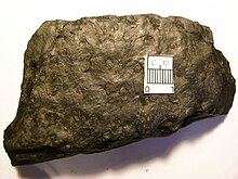
Neuropteris ovata from the late Carboniferous |
||||||||||||
| Temporal occurrence | ||||||||||||
| Lower Carboniferous to Permian | ||||||||||||
| Systematics | ||||||||||||
|
||||||||||||
| Scientific name | ||||||||||||
| Medullosales | ||||||||||||
The Medullosales are a Paleozoic order of the extinct plant group of seed ferns . They were the largest representatives of the seed ferns. The conductive tissue was divided into several segments, which were surrounded by secondary xylem .
features
The growth form was reconstructed on the basis of Alethopteris , whereby one species may have developed two growth forms depending on the environmental conditions: thin, liana-like shoot axes on which the leaf fronds sit at a greater distance, and free and upright stems on which the leaf fronds are close together.
tribe
The form genus Medullosa includes various fossils from tribes. The following species have been described for Medullosa so far : M. anglica, M. centrofilis, M. endocentrica, M. geriensis, M. gigas, M. leuckartii, M. noei, M. olseniae, M. porosa, M. pusilla, M. primaeva, M. steinii, M. stellata, M. solmsii, M. thompsonii . Furthermore, there are very different forms for some of these species .
In cross-section, the trunks are characterized by the presence of several segments of conductive tissue. In earlier work, the trunks were therefore interpreted as polysteles , today the segments are viewed as parts of a single stele.
In Medullosa noei , one of the most common species in North American Pennsylvania , the trunk contains two to four segments of conductive tissue. The number varies along an axis by branching and merging the segments. Each segment is elliptical to ribbon-shaped in cross section, the xylem contains plenty of parenchyma , the protoxylem is on the outside. Around each segment is a cylinder of secondary xylem of varying thickness. In the center of the trunk it is more developed. The tracheids of the secondary xylem are unusually long, up to 24 millimeters. There are up to twelve rows of court pits on the radial walls of the tracheids . The rays are two to eight cells wide and stand between every third row of tracheids.
Outwardly, the xylem is followed by the cambium and the secondary phloem . This is made up of alternating tangential bands of sieve cells , fibers and axial parenchyma. A cortical parenchyma with elongated secretion canals follow on the outside. The channels are a few millimeters long, filled with amorphous substance and surrounded by a layer of small epithelial cells. Often a periderm also occurs, which can be a centimeter or thicker. The periderm cells are isodiametric , thick-walled and arranged in radial rows. The structure suggests that the phellogen gave the periderm mainly inward. The peridermal tissue may have been alive and continued to divide. Both characteristics do not occur in recent seed plants.
In Medullosa primaeva the trunk has a diameter of around eight centimeters and contains two to over 20 segments.
The younger representatives from the Permian period differ in their trunk structure from the older ones: Medullosa stellata has trunks with a diameter of up to 50 centimeters and up to 43 individual central segments. Around this is a cylinder of primary and secondary xylem.
leaves
The leaf fronds of the Medullosales were very large, as far as can be reconstructed from the preserved petioles and leaf parts. The leaves are forked and evenly pinnate. The leaves are in 2/5 or 1/3 phyllotaxis on the stems. Isolated petioles of Medullosa are called myeloxylon and are up to eight inches in diameter. Inside there are scattered vascular bundles , fiber strands run close to the surface, and secretion canals are scattered in the base tissue.
The two most common genera of leaves of the Medullosales are Neuropteris and Alethopteris . In Alethopteris , the leaf margins of the leaflets are rolled down and the veins consist of one to four vascular bundles. The leaflets in Alethopteris sullivantii are, for example, 1.2 to 2.4 centimeters long and 1.2 centimeters wide. The leaflets of Neuropteris have a heart-shaped base and a distinct midrib, the vascular bundles have a parenchymal bundle sheath. In Reticulopteris muensteri , pore-like openings near the vein ends that were compared to hydathodes were discovered.
Further leaf genera of the Medullosales are Mixoneura , Linopteris , Cyclopteris , Odontopteris , Neuralethopteris , Lonchopteris , Lonchopteridium and Macralethopteris .
root
Medullosa formed adventitious roots . These are often represented in fossilized remains from the carbon. Roots on stems of Medullosa anglica are triarch and stand in vertical rows on the stem. Older roots have a thick periderm. Middle Pennsylvania representatives are up to an inch in diameter and have abundant secondary tissue. Here in the center there is an exarche actinostele with five protoxylem points. The secondary xylem is continuous here, while in other Medullosa roots the secondary xylem is only formed above the metaxylem. The secondary phloem is well formed and consists of sieve cells and rays. Lateral roots arise from a thin-walled pericycle above the protoxylemic strands. An endodermis and a parenchymal cortex are located around the pericycle .
Ovules and seeds
The seeds of the Medullosales are up to eleven centimeters in length and are among the largest among the seed ferns. In their structure they are almost identical to those of the recent cycads .
The most common genus is Pachytesta , to which around a dozen species are counted. The seed size ranges from one to eleven centimeters in length. In all representatives of the order, the seed coat consists of three equally sized valves. In Pachytesta illinoensis from North America, the seed coat (testa) consists of three layers: a parenchymal outer layer (sarcotesta), a middle layer of fibers (sclerotesta) and a single-celled innermost endotesta. The secretory ducts known from Myeloxylon and Medullosa occur in the seed coat . The Sclerotesta has three main ribs on the outside, which extend from the base to almost the micropyle. Smaller secondary and tertiary ribs can appear between these main ribs. The nucellus is only fused with the integument at the base of the ovule . At the distal end, the nucellus forms a simple bell-shaped pollen chamber. The nucellus ends in a small beak that is attached to the micropylene opening. In Pachytesta illionensis, the integument is supplied by up to 42 vascular bundles, around 25 lead into the nucellus.
Pachytesta gigantea from France and North America are seeds up to seven centimeters long, the sclerotesta of which is not ornamented with ribs. Pachytesta vera has three distinct ribs of the same size.
In some of the finds, the mega gametophytes could also be examined. In Pachytesta hexangulata , a gametophyte forms three oval archegonia with a diameter of around one millimeter. In a number of specimens, pollen grains of the genus Monoletes could be identified near the archegonia .
The seeds of Stephanospermum are around one centimeter long and have an elongated micropylene channel. They have three ribs. In Stephanospermum konopeounus there are wing-like structures on the integument. The seeds are located on branched axes, and not on leaf blades, as otherwise assumed for the Medullosales. The presence of Monoletes pollen reinforces the fact that S. konopeounus belongs to this group.
In Hexapterospermum , the sclerotesta has six ribs. The three layers of the seed wall flow smoothly into one another. The pollen chamber is simple. Rhynchospermum has a two-layer seed coat. There are eight to ten sclerotesta ribs in the upper half of the seed, while the lower half is smooth. The integument and nucellus are fused, the pollen chamber is simple.
Some genera include casts of seeds. They are placed on the medullosales because of their size and because of the ribs on the surface. They include Trigonocarpus , of which some species, such as Trigonocarpus leeanus from Middle Pennsylvania, were about 10 centimeters long. Many of the Trigonocarpus species are likely to represent casts of Pachytesta in various states of preservation. Two leaflets of Neuropteris heterophylla were found at the base of some Trigonocarpus seeds . It was concluded from this that the ovules on pinnate leaves occupied the position of the terminal leaflets. In other forms the seeds are on the midrib of a pinna. In Spermopteris , the ovules are on the underside of Taeniopteris leaf blades. In Spermopteris coriacea , the ovules are each in a row on the sides of the midrib of a leaflet, with a leaf vein leading to each ovule .
Pollen-producing organs
The pollen-producing organs are relatively large and can reach several centimeters in diameter. They are synangiate forms. Depending on the complexity, three groups can be distinguished:
- The simple, solitary forms are represented by Halletheca and Codonotheca, for example , and have separate synangia. The simplest form occurs in Halletheca reticulata : The synangium is pear-shaped and around 1.5 centimeters long. Five sporangia are arranged in a circle around a central zone of fibers. This central zone is hollow at the distal end. The vascular bundles are on the outside of the sporangia. The sporangia open towards the central zone. In Sullitheca there are around 40 sporangia in a parenchymal tissue. In the center there is an H-shaped zone made of sclerenchymal fibers similar to Halletheca .
- In the aggregate type, the synangia are grouped together, but they are not fused. Representatives are Rhetinotheca , Parasporotheca and Whittleseya . In Rhetinotheca , the single synangium is rather small at around two millimeters and consists of four sporangia that surround a central fibrous structure. The individual synangia are at the end branches of a highly branched axis system.
- The third group includes, for example, Bernaultia and Dolerotheca . In Bernaultia , the pollen organs are up to four centimeters long and are approximately bell-shaped. In Bernaultia formosa , a common species from the late Pennsylvania , the organ consists of four synangia, each strongly folded and containing numerous elongated sporangia, each opening into the cavities between the folds. There are several theories about the evolutionary development of this complex form from the simpler Halletheca -shaped organs due to the lack of intermediate forms.
In contrast to the other pollen organs, Parasporotheca is not radially symmetrical. In Parasporotheca , a synangium is spoon-shaped, the sporangia are elongated and embedded on the inside of the spoon. Several synangia stand together on branched shoot axes and are together up to 20 centimeters long.
Pollen and microgametophyte
The pollen grains are very large with a diameter of 100 to 600 micrometers. With the exception of the pollen organs Potoniea and Parasporotheca , all pollen of the genus Monoletes contains the bilateral: on the proximal side there is a monolete scar, on the distal side two long furrows over the entire length of the pollen grain. The sporoderm is you and consists of two layers. Ubisch bodies, 0.3 to 0.8 micrometers in size, associated with cell membranes have been identified on some pollen grains .
Micro gametophytes morphologically resemble those of some Cycadales representatives. Within a pollen grain there are 10 to 14 compartments, which are interpreted as cells. There is no evidence of the presence of pollen tubes.
ecology
From epidermal features of Neuropteris leaves from the Middle and Lower Carboniferous of Canada and Germany, it has been concluded that most of the species grew on humid locations in the lowlands.
There are speculations that the pollen of the Medullosales was one of the first to be spread by animals ( zoophilia ), possibly by carbon-age arthropods .
distribution
Finds of the Medullosales range from the Lower Carboniferous (beginning 359 million years ago) to the Permian (end 251 million years ago).
Systematic position
The Medullosales are one of several Paleozoic seed fern orders, which, however, are probably not closely related.
For some time they were discussed as possible precursors of the Cycadales , especially the ovules and seeds of the two groups are similar to one another. However, the pollen organs differ greatly. Cycadales fossils from the late Paleozoic are now also known, so they are the same age as the Medullosales.
Botanical history
In 1828, Adolphe Brongniart described casts of seeds as a trigonocarpus . Bernhard von Cotta described the remains of tribes from the Lower Permian as Medullosa in 1832 .
supporting documents
- Thomas N. Taylor, Edith L. Taylor: The Biology and Evolution of Fossil Plants . Prentice Hall, Englewood Cliffs 1993, ISBN 0-13-651589-4 , pp. 522-549.


