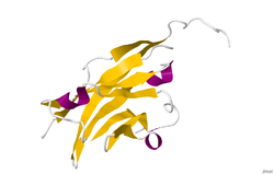
Myelin oligodendrocyte glycoprotein
| Myelin oligodendrocyte glycoprotein | ||
|---|---|---|

|
||
| X-ray crystal structure analysis of myelin oligodendrocyte glycoportein a rat according to PDB 1PKO | ||
| Properties of human protein | ||
| Mass / length primary structure | 12.1 - 33.5 kilodaltons / 108 - 295 amino acids (depending on isoform) | |
| Isoforms | 13 | |
| Identifier | ||
| Gene names | MOG BTN6; BTNL11; MOGIG2; NRCLP7 | |
| External IDs | ||
| Orthologue | ||
| human | House mouse | |
| Entrez | 4340 | 17441 |
| Ensemble | ENSG00000137345 | ENSMUSG00000076439 |
| UniProt | Q16653 | Q61885 |
| Refseq (mRNA) | NM_001008228 | NM_010814 |
| Refseq (protein) | NP_001008229 | NP_034944 |
| Gene locus | Chr 6: 29.66 - 29.67 Mb | Chr 17: 37.01 - 37.02 Mb |
| PubMed search | 4340 |
17441
|
The myelin oligodendrocyte glycoprotein (MOG) is a glycoprotein that plays an important role in the myelination process of nerves in the CNS (see also axon and conduction ). Encodes the protein in humans by being MOG - Gen . Investigations into the molecular basis of myelin formation are important for various neurological diseases in which there is a loss of the protective myelin layer, as in multiple sclerosis .
It is believed that MOG plays a role as an adhesion molecule, thereby imparting structural integrity to the myelin sheath . It arises late on the oligodendrocyte .
Molecular function
The primary molecular role of MOG is as yet unknown, but the most likely is that of completion and / or maintenance of the myelin sheath. In detail, it is assumed that MOG plays a role as an "adhesion molecule" on the myelin sheath and imparts structural integrity to the sheath . "
The cDNA coding region of MOG in humans is to a high degree homologous to rats, mice and cattle and is therefore highly conserved. This underlines the assumption that MOG plays a “biologically important role” in the organism.
physiology
The MOG gene, which is located on chromosome 6p21.3-p22, was sequenced in 1995.
It is a transmembrane protein , which on the surface of oligodendrocytes and on the outermost layer of the myelin sheath expressed is. "MOG is a type 1 transmembrane protein that occurs in small quantities and is only found in the CNS." "A single Ig domain extends into the extracellular space" and allows easy access by autoantibodies. The primary nuclear transcript of MOG comprises ... 15,561 nucleotides "and has eight exons in humans , which are separated from each other by seven introns ." The introns contain "numerous repetitive DNA" sequences, among which a "14 Alu sequence within 3 Introns are located. ”The length of the introns varies between 242 and 6484 base pairs.
structure
Due to alternative splicings of the human mRNA, at least nine isoforms result from the MOG gene.
The crystal structure of the myelin oligodendrocyte glycoprotein was determined using the protein from the traveling rat by means of X-ray crystal structure analysis with a resolution of 1.45 Angstroms. The protein comprises 139 amino acids and belongs to the immunoglobulin superfamily .
The DSSP secondary structure (Define Secondary Structure of Proteins) of the protein consists of 6% helices and 43% of β-sheet : Three short helical segments followed by ten β-sheets. The β-sheet sequences lie within two antiparallel β-sheets which form an immunoglobulin-like β-sandwich fold. Several properties of the protein structure of the MOG indicate a role as “adhesin for completing and / or compressing the myelin sheath.” Starting near the N-terminus, there is a strip of electronegative charge that extends over half the length of the molecule. In solution, MOG shows the tendency towards dimerization and the "shape complementary index" is high at the dimerization junction, suggesting the existence of a "biologically relevant MOG dimer".
synthesis
MOG is only formed at a “relatively late point in time” on the oligodendrocytes and the mycelial envelope.
Diseases
The main interest in MOG lies in its role in connection with demyelinating diseases such as B. adrenoleukodystrophy , childhood ataxia with central myelination , multiple sclerosis (MS) and rubella- induced intellectual disability .
MOG is a target antigen that leads to autoimmune demyelination . Most of the scientific laboratory studies on MOG are related to MS. Various studies have shown a connection between MOG antibodies and the pathogenesis in MS. Animal models of the MS EAE showed (in different animal lines) that this MOG-specific EAE is similar to the MS or almost entirely "copies" it. as evidenced by the demyelinating capacity and topography of the lesions . The animal models were able to show that MOG antibodies are responsible for demyelination. These models have been studied extensively and MOG are the only antibodies so far that have the ability to trigger demyelination.
multiple sclerosis
The exact pathogenic processes of MS are so far unknown, however, based on the current state of knowledge, various theories exist: The theory of antibody-mediated demyelination , an attack by the immune system on the body, in particular on the central nervous system , which ultimately leads to the Demyelination leads. According to this theory, specific antigens in the body are targeted. In detail, T and B cells are held responsible as the cells leading to pathogenesis in the antibody-mediated demyelination of MS. Among the various candidates who may be involved in the pathogenesis, the scientific focus in the studies mentioned below is mostly on two antigens: One is myelin basic protein (MBP), in which various antibodies directed against MBP could be detected in the early stages of MS. The other is myelin oligodendrocyte glycoprotein (MOG). Both were identified as "targets of the immune response". In connection with the "antibody-mediating demyelination theory", these antibodies, which arise during the immune response, could play an important role in the development of multiple sclerosis.
One study found a correlation between MS relapses and anti-MOG and anti-MBP antibodies in the blood serum of patients with a " clinically isolated syndrome "; these relapses ultimately led to the definitive clinical diagnosis of MS in these patients. Patients who were seronegative for both antibodies suffered a relapse in only 23% of all cases after 45.1 ± 13.7 months, whereas 83% of the patients who tested positive for anti-MOG antibodies alone within 14.6 ± 9, Had a relapse for 6 months. 21 of 22 patients who had both antibodies in their blood had relapses within a period of 7.5 ± 4.4 months.
These results suggest that the occurrence of a clinically isolated syndrome does not necessarily have to lead to "clinically definitive" MS. These results are consistent with previous data on the disease. For example, 30-40 percent of MS cases have a relatively harmless disease course, which correlates with the 38 percent of patients in this study who were negative for both antibodies. In the early course of the disease, the “antibody status” seems to offer a way of identifying patients with a likely milder course of the disease. The article also emphasizes that these results do not demonstrate that the antibodies lead to demyelination. Rather, they provide a useful diagnostic indicator of the future course of the disease. A possible practical application of the study would be a cheaper and simpler alternative to the currently common “ Magnetic Resonance Imaging ” strategy for determining the risk of relapse after a “clinically isolated syndrome”.
A comparable study examined the change in the risk (depending on the antibody status) of developing clinically definitive MS after a previous clinically isolated syndrome. The anti-myelin antibodies mentioned were examined as a possible predictor of a changed risk. While 90 percent of patients with clinically isolated syndrome were generally diagnosed with clinically definitive MS within months to years, the results of the study also showed that patients with a negative test for antibodies generally had a better prognosis at the time of relapse (ie relapse generally occurred later). In return, patients who tested positive for the antibodies were able to benefit from starting therapy as early as possible due to the test. Over a period of 12 months, 30 patients tested positive for the antibodies. 22 of these patients developed clinically definitive MS. In contrast, none of the patients who were negative for the antibody developed clinically definitive MS within the period mentioned.
Despite these results, another work concluded that the studies mentioned could not conclusively show that MOG is in fact one of the main contributors to the course of MS. MOG has been shown to be able to cause demyelination in vitro and in experimental animal models. It can also be detected in nerve tissue lesions as well as in patients with diagnosed MS. However, the significance of these discoveries is not yet clearly conclusive. Two other studies were only able to confirm the results from the studies mentioned in a subgroup analysis and three further studies on the topic came to negative results. The work mentioned offers a different interpretation of the results: It states that the anti-MOG antibody correlation to the development of MS may at least partially reflect the cross-reactivity between MOG and the nuclear antigen of the Epstein-Barr virus (EBNA). The association of MOGs with MS arose from its synthesis site, which is exclusively in the central nervous system. However, the true relationship between MS and MOG is not fully understood, partly due to the lack of evidence on the relationship between biologically active anti-MOG antibodies and the demyelination that ultimately leads to MS. Finally, while anti-MOG antibodies can be measured to determine the extent of tissue damage during MS, other than biologically active antibodies, they could also simply be an accompanying phenomenon of CNS tissue degeneration.
Web links
Individual evidence
- ↑ Pham-Dinh D, Della Gaspera B, Kerlero de Rosbo N, Dautigny A: Structure of the human myelin / oligodendrocyte glycoprotein gene and multiple alternative spliced isoforms . In: Genomics . 29, No. 2, September 1995, pp. 345-52. doi : 10.1006 / geno.1995.9995 . PMID 8666381 .
- ↑ a b Pham-Dinh D, Jones EP, Pitiot G, Della Gaspera B, Daubas P, Mallet J, Le Paslier D, Fischer Lindahl K, Dautigny A: Physical mapping of the human and mouse MOG gene at the distal end of the MHC class Ib region . In: Immunogenetics . 42, No. 5, 1995, pp. 386-91. PMID 7590972 .
- ↑ a b c d e f g h Roth MP, Malfroy L, Offer C, Sevin J, Enault G, Borot N, Pontarotti P, Coppin H: The human myelin oligodendrocyte glycoprotein (MOG) gene: complete nucleotide sequence and structural characterization . In: Genomics . 28, No. 2, July 1995, pp. 241-50. doi : 10.1006 / geno.1995.1137 . PMID 8530032 .
- ↑ a b c d e f g h i Berger, T., Innsbruck Medical University Dept. of Neurology interviewed by S. Gillooly, Nov. 24, 2008.
- ↑ Danielle Pham ‐ Dinh, Bernadette Allinquant, Merle Ruberg, Bruno Della Gaspera, Jean ‐ Louis Nussbaum, André Dautigny : Characterization and Expression of the cDNA Coding for the Human Myelin / Oligodendrocyte Glycoprotein . In: John Wiley & Sons (Eds.): Journal of Neurochemistry . Volume 63, Issue 6 , December 1994, pp. 2353-2356 , doi : 10.1046 / j.1471-4159.1994.63062353.x (English).
- ↑ Pham-Dinh D, Mattei MG, Nussbaum JL, Roussel G, Pontarotti P, Roeckel N, Mather IH, Artzt K, Lindahl KF, Dautigny A: Myelin / oligodendrocyte glycoprotein is a member of a subset of the immunoglobulin superfamily encoded within the major histocompatibility complex . In: Proc. Natl. Acad. Sci. USA . 90, No. 17, September 1993, pp. 7990-4. doi : 10.1073 / pnas.90.17.7990 . PMID 8367453 .
- ↑ a b c Berger T, Reindl M: Multiple sclerosis: disease biomarkers as indicated by pathophysiology . In: J. Neurol. Sci. . 259, No. 1-2, August 2007, pp. 21-6. doi : 10.1016 / j.jns.2006.05.070 . PMID 17367811 .
- ↑ Boyle LH, Traherne JA, Plotnek G, Ward R, Trowsdale J: Splice variation in the cytoplasmic domains of myelin oligodendrocyte glycoprotein affects its cellular localization and transport . In: J. Neurochem. . 102, No. 6, September 2007, pp. 1853-1862. doi : 10.1111 / j.1471-4159.2007.04687.x . PMID 17573820 .
- ↑ Breithaupt C, Schubart A, Zander H, et al. : Structural insights into the antigenicity of myelin oligodendrocyte glycoprotein . In: Proc. Natl. Acad. Sci. USA . 100, No. 16, August 2003, pp. 9446-51. doi : 10.1073 / pnas.1133443100 . PMID 12874380 .
- ↑ Kabsch W, Sander C: Dictionary of protein secondary structure: pattern recognition of hydrogen-bonded and geometrical features . In: Biopolymers . 22, No. 12, December 1983, pp. 2577-637. doi : 10.1002 / bip.360221211 . PMID 6667333 .
- ↑ Murzin AG, Brenner SE, Hubbard T, Chothia C: SCOP: a structural classification of proteins database for the investigation of sequences and structures . In: J. Mol. Biol. . 247, No. 4, April 1995, pp. 536-40. doi : 10.1016 / S0022-2836 (05) 80134-2 . PMID 7723011 .
- ↑ Clements CS, Reid HH, Beddoe T, et al. : The crystal structure of myelin oligodendrocyte glycoprotein, a key autoantigen in multiple sclerosis . In: Proc. Natl. Acad. Sci. USA . 100, No. 19, September 2003, pp. 11059-64. doi : 10.1073 / pnas.1833158100 . PMID 12960396 .
- ↑ Cong H, Jiang Y, Tien P: Identification of the myelin oligodendrocyte glycoprotein as a cellular receptor for rubella virus . In: J. Virol. . 85, No. 21, November 2011, pp. 11038-47. doi : 10.1128 / JVI.05398-11 . PMID 21880773 .
- ↑ a b c d e Berger T, Rubner P, Schautzer F, Egg R, Ulmer H, Mayringer I, Dilitz E, Deisenhammer F, Reindl M: Antimyelin antibodies as a predictor of clinically definite multiple sclerosis after a first demyelinating event . In: N. Engl. J. Med. . 349, No. 2, July 2003, pp. 139-45. doi : 10.1056 / NEJMoa022328 . PMID 12853586 .
- ↑ a b c Tomassini V, De Giglio L, Reindl M, Russo P, Pestalozza I, Pantano P, Berger T, Pozzilli C: Anti-myelin antibodies predict the clinical outcome after a first episode suggestive of MS . In: Mult. Scler. . 13, No. 9, November 2007, pp. 1086-1094. doi : 10.1177 / 1352458507077622 . PMID 17468447 .
- ↑ a b Greeve I, J Sellner, Lauterburg T, U Walker, Rösler KM, Mattle HP: Anti-myelin antibodies in clinically isolated syndrome indicate the risk of multiple sclerosis in a Swiss cohort . In: Acta Neurol. Scand. . 116, No. 4, October 2007, pp. 207-10. doi : 10.1111 / j.1600-0404.2007.00872.x . PMID 17824895 .
- ↑ a b Wang H, Munger KL, Reindl M, O'Reilly EJ, Levin LI, Berger T, Ascherio A: Myelin oligodendrocyte glycoprotein antibodies and multiple sclerosis in healthy young adults . In: Neurology . 71, No. 15, October 2008, pp. 1142-6. doi : 10.1212 / 01.wnl.0000316195.52001.e1 . PMID 18753473 .