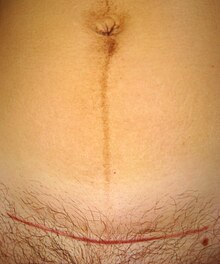
Scar (wound healing)
| Classification according to ICD-10 | |
|---|---|
| L90.5 | Scars and fibroses of the skin |
| ICD-10 online (WHO version 2019) | |
As scar ( latin Cicatrix ) of the after destruction of the collagenous network of the skin , a fiber-rich replacement tissue ( fibrosis hereinafter), the final state of a wound healing represents. In scars, the collagen is no longer complexly intertwined, but rather arranged in parallel. There are no skin appendages such as sebum or sweat glands .
In the case of traumatic and different lesions with severing or loss of tissue, the wound is first closed with a fibrin clot , then inflammatory cells are recruited. Granulation tissue then forms , which is finally replaced by collagen-rich connective tissue , which tightens as the healing process continues. In addition, there is a re-epithelialization of the skin surface.
Complications during wound healing negatively affect healing. With regard to conspicuous scar formation, a distinction can be made between atrophic scars, scar contractures, hypertrophic scars and keloids .
skin
Superficial lesions of the epidermis heal without consequences in the sense of a “ restitutio ad integrum ”. Such superficial wounds, caused by abrasion or sunburn, heal in the regenerative process of epithelial wound healing without scars and the damaged tissue is restored within a few days. When the skin is injured, a scar only occurs if the dermis has been injured. Because the scar tissue is initially crossed by many blood vessels ( angiogenesis ), it has a red color. As the remodeling continues, the blood vessels recede, while the proportion of collagen fibers increases. On the one hand, this increases the tear resistance of the scar; on the other hand, it now becomes lighter than the surrounding tissue, since the melanocytes are missing in the scar tissue (at least initially, but possibly permanently). Despite the large number of collagen fibers, a scar is an inferior tissue. When exposed to heavy loads, scar hernias can occur later in life .
While melanocytes can immigrate again, remnants of hair follicles , sebum or sweat glands with their epithelial tissue initially play an important role in the re-epithelialization of the skin surface. If these remnants have gone under, they will not be re-formed and will be permanently missing in a scar.
Special forms are the keloid and the hypertrophic scar .
Scars from other organs
Even if other organs are damaged, their dead tissue-typical cells are replaced by scarred connective tissue. After a heart attack , the heart muscle cells die . The size of the scar that occurs causes the heart to pump less and can lead to arrhythmias . After pyelonephritis , permanent scarring can occur in the kidneys if it is treated with medication too late. Kidney scars can cause high blood pressure. A corneal scar (such as a leukoma) can appear on the eye.
Scar treatment
Good wound care while the wound is healing can have a positive effect on appearance and functionality, but cannot completely prevent it. So-called wound closure plasters and tissue adhesives are used for primary and secondary healing wounds, protect the wound edges from damage and trauma and enable pain-free wound closure with little scarring. The skin adhesives used here are highly viscous and relatively easy to apply.
Since the fresh scar lacks melanocytes, it produces less melanin than the surrounding skin and is therefore sensitive to UV radiation.
A subsequent scar treatment that would completely eliminate scars is still not possible today. Excessive scars can be improved by means of scar mobilization or invasive procedures (such as laser, surgery, nitrogen freezing or dermabrasion ); however, there are risks of new scars in the surgical procedures.
Scars can also be treated by massaging in special scar creams ( e.g. gels containing silicone or a combination of heparin , allantoin and onion extract) several times a day for months ; ultrasound is also used to support the effectiveness of scar gels containing heparin . Scar plasters (especially silicone pads, especially for hypertrophic scars, or adhesive tapes with micropores) are also used to reduce scar bulges and burns. There are contradicting results on the effectiveness of these procedures.
In the case of large-scale injuries (such as burns), compression bandages that are to be worn for months or years are used.
See also
- Secondary healing - wound healing with severe scarring
- Contracture - permanent shortening of muscles due to extensive scars (dermatogenic contracture)
- Scarification - decorative scars as body jewelry
- Pleural rind
- Schmiss - the scar of a student scale acquired by being hit by a cutting weapon
- Glasgow Smile
- Moist wound treatment and dry wound treatment
literature
- Kerstin Protz: Modern wound care. 7th edition. Elsevier Urban & Fischer, Munich 2014, ISBN 978-3-437-27884-6 .
Web links
- Skin Injuries: What Helps With Scars , Spiegel Online, March 22, 2017
Individual evidence
- ↑ Konrad Herrmann, Ute Trinkkeller: Dermatology and Medical Cosmetics: Guide for Cosmetic Practice. Springer-Verlag, 2007, ISBN 978-3-540-30094-6 , p. 141.
- ↑ Hans Lippert (Ed.): Wundatlas Compendium of complex wound treatment. Georg Thieme Verlag, Stuttgart, 2006, ISBN 3-13-140832-4 , p. 31.
- ↑ Bernd von Hallern: Compendium Wound Treatment Mode of action and indication of wound treatment products. Publishing house for medical publications, Stade 2015, page 115.
- ↑ Alan Widgerow: Scar management - marrying the practical with the science. In: Wound Healing Southern Africa. Volume 3, No. 1, 2010, pp. 7-10.
- ↑ Alain Widgerow among others :: multimodality scar management program. In: Aesthetic Plast Surg. Volume 33, No. 4, Jul 2009, pp. 533-543. PMID 19048338 .

