Optical spectrometer
An optical spectrometer is a spectrometer for visible light and adjacent areas of the electromagnetic spectrum . It can be used to obtain emission spectra (spectral examinations of light sources) as well as absorption spectra and statements on frequency-dependent reflection .
Structure of a prism or grating spectrometer
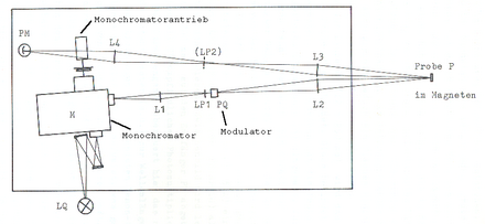
The following setup is typical for a grating spectrometer in the VIS or NIR range:
A light source LQ, depending on the wavelength range z. B .:
- Xenon high-pressure lamp ( gas discharge lamp , visible spectral range and adjacent areas)
- Halogen lamp (visible up to MIR)
- Tunable lasers (imaging optics and monochromator can be omitted)
An imaging system (two mirrors in the picture) images the light source LQ onto the monochromator entrance slit . An adjustable monochromator M is used to set the wavelength that passes through . He is z. B. driven by a stepper motor and also provides the value of the wavelength for evaluation.
Another imaging system L1, L2 focuses the radiation from the monochromator exit slit onto the sample.
The sample P to be examined is, for example, a reflector in the image. In other cases a sample chamber ( cuvette ) is irradiated or the light source itself is the object to be examined.
With the imaging system L3, L4, the signal is imaged on a photoreceiver PM. Possible receivers PM (see also radiation detector ) are:
- Photodiodes and semiconductor detectors for the visible and adjacent areas and - with appropriate cooling - also into the mid-infrared (MIR)
- Photo multiplier (PM) for the visible range and ultraviolet
- Bolometers and pyroelectric sensors in the middle and far infrared
A registration and analysis system combines the current values of the monochromator wavelength and the receiver signal, displays them in a measurement curve and analyzes them. Today this is usually a computer with the appropriate interfaces plus software .
Compact spectrometer
The aim is to achieve compact devices that can easily be used in any application. All moving parts should be dispensed with, which greatly reduces the required size and improves the reproducibility of the measurements. This is achieved by receiving and evaluating the light, broken down into colors, by a line of photodiodes , whereby each individual sensor is there for a certain wavelength range (always the same). The measured values for the entire spectrum are therefore available in parallel on the individual sensors. Parts of the imaging optics are sometimes designed as fiber optics .
This has certain consequences for the structure:
- Since the beam for each wavelength runs differently behind the dispersing element (still prism or grating), a sample to be measured must be inserted into the beam path in front of this element - for example a liquid in a cuvette . Furthermore, a design for both transmission and reflection is possible.
- However, more complicated elements in the beam path, such as certain modulators or high field magnets or cryostats , can hardly be used here.
- The primary light source is also integrated. Optionally, it can be flange-mounted as an exchangeable module.
- The evaluation system has to be adapted, but it is more simple than more complicated.
- The entire device can be designed as a compact, hermetically sealed box (with access to a sample lock if necessary).
FT spectrometer
FT spectrometers work on the principle of an interferometer , in which the signal is evaluated with the aid of the Fourier transformation (FT) with regard to the frequencies it contains while the interferometer is being adjusted. The main advantage of the FT spectrometer is the shorter measurement time, since in contrast to dispersive systems (prism or grating spectrometers) the sample does not have to be irradiated step by step with a changing frequency. These spectrometers are mainly used in the infrared range ( see also: FTIR spectrometers ), but FT spectrometers for other spectroscopic methods such as Raman spectroscopy are also available on the market .
variants
In certain investigations into photoconductivity , the sample itself forms the receiver, so that one of the imaging systems and the photo receiver are not required.
In MIR and ultraviolet from about 200 nm, the images must be made with concave mirrors (e.g. aluminum on glass), since glass is no longer transparent. Mirrors also have the advantage of a wavelength-independent imaging geometry, while lenses without adjustment can only be used for a narrow spectral range.
A modulator is often arranged between the light source and the monochromator in order to be able to better distinguish the signal from the ambient light when evaluating the receiver signal. The modulator can e.g. B. be a polarization modulator or a simple chopper disk.
There are also spectrometers with a polychromator that do not scan the spectrum sequentially, but record it simultaneously. The dispersing or refractive element is only arranged behind the sample and the spectrum is received simultaneously by a line camera , i.e. a linear arrangement of photodiodes, so that the evaluation electronics only have to query and register this row of receivers. See also diode array detector .
Echelle polychromators use area detectors to evaluate the spectrum.
Applications
Optical spectrometers are mainly used for solid-state spectroscopy:
- Reflection spectra are recorded by measuring a spectrum of the reflectance with the sample and then a spectrum in which the sample is replaced by a reference mirror with a known reflection spectrum. A suitable reference mirror material for visible light and infrared is aluminum (vapor-deposited on glass), which achieves a degree of reflection of close to 1 in this wavelength range without strong structuring.
- Transmission or absorption spectra are recorded by introducing the material to be examined into the beam path at the location of an intermediate image. This spectrum is then compared with a reference spectrum without any sample.
- With photoconductivity spectra , the sample is used as a receiver. As a reference, you have to replace the sample with a receiver with a known spectral response.
At least the absorption measurements can also be carried out on liquids and, in extreme cases, on gases using a cuvette .
Depending on the details of the question, various optical modulators are used to obtain an alternating light signal that specifically addresses certain ( e.g. magneto-optical ) properties of the sample and which can be better processed as an electrical signal after the receiver (e.g. using a lock-in amplifier ).
Technical implementation
Individual evidence
- ↑ Patent WO2004070329A2 : Compact spectrometer. Published on August 19, 2004 ( PDF file, 5.3 kB ).
- ↑ Spectroscopy with compact spectrometers ( Memento of the original from March 7, 2014 in the Internet Archive ) Info: The archive link was inserted automatically and has not yet been checked. Please check the original and archive link according to the instructions and then remove this notice. (Company publication, PDF file, 1.3 MB)
