Six-point thief beetle
| Six-point thief beetle | ||||||||||
|---|---|---|---|---|---|---|---|---|---|---|
![Illustration by John Curtis from 1887 [1]](https://upload.wikimedia.org/wikipedia/commons/thumb/a/a8/Britishentomologyvolume2Plate646_retusche_2.jpg/225px-Britishentomologyvolume2Plate646_retusche_2.jpg)
Illustration by John Curtis from 1887 |
||||||||||
| Systematics | ||||||||||
|
||||||||||
| Scientific name | ||||||||||
| Ptinus sexpunctatus | ||||||||||
| Tank , 1789 |
The six-point thief beetle ( Ptinus sexpunctatus ) is a beetle from the rodent beetle family . The genus Ptinus is represented in Europe with six sub-genera, Ptinus sexpunctatus is included in the sub-genus Gynopterus , which is represented in Europe with nine species.
The species is classified as endangered (category 3) in the Red List of Endangered Animals, Plants and Fungi in Germany .
Notes on the names
The first description is occasionally incorrectly given as Panzer 1795 . In the Entomological Pocket Book for the year 1795 by Panzer - the preface is dated 1794 - Panzer mentions himself twice as the first person to describe the Beetle. The one place he cited was not published until the following year, 1796. Here the description of the beetle includes a color plate that shows the beetle in natural size and enlarged and is signed Ptinus sexpunctatus Mihi ( Latin mihi - "me, my": tank claims the beetle's authorship). The color table is followed by a short Latin description and some German comments on the beetle, including information on the habitat in dark, dusty places in old buildings and Panzers' reference to an earlier description of the beetle from his pen. This first description appeared in 1789, is in Latin and is relatively detailed for the time.
On each wing cover of the beetle one can see two white spots on superficial inspection, so a total of four spots, which seems to contradict the species name sexpunctata (Latin "with six points"). In Panzer's first description, however, with regard to the stains, it is stated that solitario ad baseos marginem, et duobus versus apicem (with a single (stain) at the edge of the base and two towards the end (of each wing cover)). Reitter formulates in this regard with two large, white, scaly transverse spots, the rear one usually appearing to be broken up into two spots . The German part of the name six points is the translation of the Latin species name sexpunctatus into German. Panzer himself calls the beetle drill beetle with six points .
The genus Ptīnus was established by Linné in 1767 as the 192nd genus. From the description of the genus it is not clear what the generic name refers to. According to Schenkling , he is from old Gr. πτηνός "ptenós" for "feathered" derived and justified by the fact that the beetle Ptilinus pectinicornis , which is noticeable for its feathery antennae, was counted by Linnaeus to the genus Ptinus .
The name of the subgenus Gynópterus is after Schenkling from old Gr . γύνε gynē for "woman" and πτερόν pterón for "wing" derived and refers to the fact that the wing covers differ in males and females. However, this does not apply to the six-point thief beetle.
The species names basicornis (from Latin basālis - "distinguished by the base" and cornis - "antennae", for the antennae thickened at the base), dispar (from Latin díspar - "unequal, different"), gavoyi (after the entomologist Louis Gavoy from Carcassonne ) and massiliensis (for a variant found near Marseille (Massīlia)) are used as synonyms.
Properties of the beetle
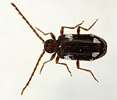
The beetle becomes 2.8 to 4.2 millimeters long. It is dark brown and has white spots on the wing covers. The antennae and legs are lighter brown. There are also red-brown colored animals.
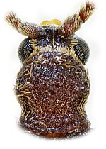
The short head is tilted downwards. Measured over the strongly bulging eyes, it is slightly narrower than the pronotum (Fig. 6) and is worn in such a way that when viewed from above it is hidden under the pronotum. The forehead is dense white scales. The eleven-link antennae are thread-shaped. They are hairy and curled up close together between the cheeks. In the male they are about body length, in the female by a quarter shorter, the eighth to tenth limb in the male about three times as long as they are wide, in the female only twice as long as they are wide. In both sexes, the first antenna segment is curved and longer than the second. The shape of the individual sensor elements is shown in Fig. 4 on the right.
The upper lip (Fig. 4, Fig. 1) is wider than it is long and ends with only a very weak margin at the front. It is hairy long on the front edge and on the sides and curved towards the middle. The strong upper jaws (mandibles, Fig. 4, Fig. 2) are hairy on the outside. They have a pointed end, have a tooth on the inner edge in the upper half and are very finely haired on the inner edge below. The hardening of the tip of the mandible with metals, which is common in insects, occurs in the genus Ptinus through the storage of zinc.
The last link of the four-link jaw probe (Fig. 4, Fig. 3 tinted blue) is as long as the three previous ones together, thickened in the middle and rounded at the tip. The end link of the short three-part lip switch (Fig. 4, Fig. 4 right tinted green) is pear-shaped.
The pronotum is about the same length as it is wide and protrudes above the head in front. Halfway up on each side, it has two weak, elongated humps parallel to each other. The sparse but strong light brown bristle hairs emphasize the course of the humps and form a vortex or tuft at their highest point. In front of the base, the pronotum is constricted in a ring shape, the constriction is not interrupted laterally by longitudinal calluses (Fig. 5).
The label is clearly visible and appears strikingly white because of its hair.
The elytra are together significantly wider than the pronotum. They are about twice as long as together wide. You have rounded, but clearly developed shoulders, the sides run almost parallel to each other behind the shoulders, in the last third the wing-coverts end together rounded. In places they have narrow, almost white, elliptical scales (Fig. 2). These form a large frayed spot behind the shoulders. In addition, they are close to the end of the wing-coverts, forming two separate, smaller, closely spaced spots, which can also be united to form a transverse band. The wing covers have ten longitudinal rows of large, densely packed, elongated rectangular points , the length of which is about the same as the width of the intervals between the point strips. On each of the intervals there is a row of moderately long, light brown bristle hairs rising diagonally backwards (Fig. 5).
The metasternum is wider in the male than in the female, a center line extends from the base to the center of the sternum, in the female this center line is only formed in the front third of the metasternum.
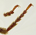
The legs are long and slender, all the tarsi are five-limbed. The fourth tarsal segment is partially lobed, cut out on top and no narrower than the third segment (Fig. 3).
Larva and pupa
The soft, fleshy, dirty white larva becomes four to five millimeters long and a good millimeter wide in the last stage with a normal, curved posture. Figure 5 on the left gives a rough idea. When stretched, the larva becomes a little longer. The larva consists of the head and twelve segments, which appear divided by longitudinal and transverse bulges (Fig. 8, Fig. 1). With the exception of the detached head, the body widens backwards without any abrupt change. He is thickly haired. The underside is flattened, the end of the body bluntly rounded.
The head yellowish due to the chitinization (Fig. 8, Fig. 2 from the front, Fig. 3 from below) is wider than long, slightly arched, and ends broadly truncated in front. He has long red hair. A center line can be seen on the head skeleton, with a characteristic large orange, elongated spot running on both sides. The center line forks indistinctly towards the rear, the two branches run out over the base of the antennae.
Above the mouth opening is a dark pigmented area below the surface, which, according to Xambeu, is divided into two spots. It is also visible through the yellowish upper lip. The edge of the upper lip is densely short reddish eyelashes. The strong upper jaws (Fig. 8, deeply dark in Fig. 2) are short, triangular and curved inward. The base is reddish, the tip black and bluntly pointed. There is a small elongated pit on the outside of the upper jaw and a pointed tooth on the inside.
The lower jaws are fleshy, reddish and hairy. The ark (Fig. 8, Fig. 3 green) is small and lined with hairs on the inside. It is only slightly surmounted by the three-part, yellowish jaw palpation (Fig. 8, Fig. 3 blue). The base link of the jaw probe is approximately ring-shaped, the second link a little longer and straighter, the end link slender, pointed and slightly curved inward. The two-part lip probes (Fig. 8, Fig. 3 red) are very short, reddish, and sit on the hairy, fleshy lower lip of the same color. The very short tripartite antennae (Fig. 8, Fig. 7 left from above, right from the side) arise behind the middle of the base of the upper jaw and point downwards. The base part of the whitish antennae is hump-like, the two following parts are very small and can be turned out. There is a single eye near the lower corner of the mandible base. The size and location of the individual parts of the head skeleton can be seen from Figures two and three of Figure eight.
The rear edge of the head is in the much larger first body segment. The first three body segments (chest segments) have long, reddish hairs, the following abdominal segments have hair of the same color, but somewhat sparse, at the end of the body the hair becomes stronger again. The breast segments each have a pair of five-limbed legs that are far apart. These (Fig. 8, Fig. 5) are only weakly developed and also hairy. They end in a short claw with an inwardly curved tip and a bristle hair.
The body segments are clearly separated only on the underside. On the upper side, with the exception of the last two segments, they are deeply incised, creating transverse ridges that pull down on the sides. The incisions and ridges are increasingly weaker towards the rear, but are particularly strong in the chest segments; in the first segment, the rear ridge even covers the transverse incision in the back area. In addition, in the rear part of the body, an elongated side bulge is formed laterally under the transverse bulges per segment, which is particularly pronounced on the last abdominal segment (anal segment).
The rounded respiratory openings (stigmas) are small and yellow with a reddish border. One stigma on each side is located on the first eight abdominal segments in the first third above the side bulge, another pair of stigmas is located near the dividing line between the first and second breast segments below the side bulge.
The underside is sparsely hairy and flattened, the separation of the individual segments clearly visible. The large anal segment is widened and carries an inclined longitudinal anal slit. At the end there is a horseshoe-shaped spot (Fig. 8, Fig. 4). As already mentioned, a strongly bulging bead runs along the sides of the anal segment, which has an important function in locomotion.
By excreting the material for the cocoon, the larva loses volume; the pupa is four millimeters long and 1.5 millimeters wide and is somewhat shorter and slimmer than the larva. An approximate picture of the view of the underside of the doll is shown in Figure 5 on the right.
biology
The heat-loving beetles can be found in very different places. On the one hand, finds in the rotten wood of old deciduous trees of different genera, especially in rot-rotten stumps of oaks , also under dry bark or in the moss on the trunks of different deciduous trees are mentioned. Finds of pines are also reported from Sweden. According to Calwer, the beetle can be found under maple bark , in cellars and in old bee and bird nests . Especially the nests are to birds' nests House Sparrow and House Martin called. A report of the meeting of the French Entomological Society explains why and how the beetle can often be found in sand pits and even observed while mating. Often one finds the imagines in the interior or near nests of Hymenoptera . Mason bees ( Osmia bicornis and other species of the genus Osmia and Chalicodoma muraria ), leafcutter bees of the genus Megachile , scissor bees (genus Chelostoma ) and hoplitis adunca are mentioned in particular . According to Müller, the beetle can be found in the wax waste of dead beehives or broken wax tablets that are left undisturbed for a long time somewhere in the apiary , apparently the honeybee is being talked about . Wasp nests, beehives and anthills are reported to be found in Spain . Especially in the northern areas of the distribution area (England, Sweden) the beetles are not infrequently found in houses. They were also found in flour and by-products of flour extraction.
These diverse habitats can be explained by the fact that the beetle feeds on remains of dead insects, but also on remains of the collecting activity of bees, and the larvae mainly develop in the nests of hymenoptera. Regarding the question, which has not yet been finally clarified, whether the larva damages the bees' nests in certain cases, Nicolas remarks: Here (in Ptinus sexpunctatus ) you can see the brazen parasite, the cheeky hoarder replaced by a renowned economist, a hardworking user as well as a clever one Calculator (from fr. On le voit l'audaxieux parasite, l'accapareur effronté est ici remplacé par un économiste rené, c'est un usager laborieux autant que prudent calculateur ). Nicolas refers to the fact that, in contrast to some parasitic beetles, according to his observations, the eggs of Ptinus sexpunctatus are only deposited in already abandoned nests and the adults only nibble on the supplies. This also explains that the rather hidden beetle can be found more easily in spring when it visits the bees' houses to lay eggs.
This opinion is being put into perspective today. Ball beetles ( Ptinus sexpunctatus ) occur occasionally in the nesting aids of mason bees. They probably lay their eggs in the open cells during the provisioning phase. The beetle larvae eat pollen and bee droppings, but it has also been observed that bees (whether larvae or imagines, dead or living specimens remain unclear) were eaten .... Since the ball beetles occur only rarely and do not tend to multiply, they are no great danger for mason beekeeping. Nevertheless, they should be removed regularly ... In order to destroy the offspring of the ball beetles, the nesting aids are opened in autumn, intact bee cocoons are removed and the ball beetle larvae are destroyed with the subsequent cleaning of the nesting aids.
In connection with parasites of Osmia cornuta , it has also been proven that the beetle at least also uses inhabited brood cells. The lid of the brood cells is either damaged by the hatching bee, then only a small wreath of mortar remains. Or the damage is in a centrally located two millimeter hole that is blamed on Ptinus sexpunctatus . Or you can find smaller holes on the periphery that are drilled by parasites.
In view of the different types of bees with which the beetle nestles as commensals , it is to be expected that it will show flexible behavior. Nicolas describes his observations in connection with the mason bee Osmia cornuta , which creates up to 24 brood cells on top of each other when developing in reed stalks, each separated by a layer of clay. Only after the fully developed bees have left the reed tube by successively gnawing through the partitions do the female beetles penetrate and lay their eggs. The larvae feed on the remains that they find in the brood cells. These are primarily the remains of the food that was provided by the bee for the development of its larva, and secondly the moulting remains and the excrement of the bee larva. To pupate, the beetle larvae gnaw a shallow hollow in the inner wall of the reed, and glue the resulting nail with vomit as a vault over the hollow. The result is a slightly curved housing, four to five millimeters long and three millimeters thick at the widest point. Nicolas describes the cocoon poetically: It is a trench spanned by a vault, built with glued material that the larva dug underneath. The house rises up where the stones were broken, at the point of extraction, the coarsest fragments on the outside, the inside is lined. It is not that the larva displays great craftsmanship, the inside of the cradle is very rough, sometimes uneven. The white silk that lines it, poorly prepared, not evenly distributed. It is an irregularly applied plaster that covers the walls, the planning leaves a lot to be desired. But the clever way the larva shows, using what is available to it, may very well outweigh the small disadvantages in architecture as an advantage (somewhat shortened from the French). In the egg-shaped cocoon thus formed, the transformation to the finished insect takes place. After curing, the imago bites a round hole in a cap of the cocoon. The first beetles appeared in mid-March, and in April they all left the cocoon. In the wild, Nicolas found an average of six beetles per opened reed that was previously used by bees for reproduction. During breeding he found up to four eggs of the beetle in the individual abandoned brood cells.
In order to investigate the flexibility of the behavior during the construction of the cocoon, Nicolas put adult beetles in a clean jar in April and provided them with plenty of pollen cake from the bees' brood cells as food. The beetles ate the food offered without haste. The droppings were deposited in long, thin, gold-colored threads, which after a while covered the entire glass floor. In mid-August there were numerous larvae on the glass floor under the excrement, which were covered by the thread-like excrement and fragments of the adults, who have now partially died. The first pupae were found in September, and after two weeks the majority had pupated and some adults had hatched. But larvae and pupae were still present at the end of May of the following year. The larvae did not have any plant material to make the dolls' cradles; they built the cocoons from the excrement of the larvae and adults as well as the remains of the dead adult animals. The coarsest pieces were outside, cleverly built in but not concealed. Inside the cocoon, the smaller fragments and fine dust provide the material that the larvae stick together with vomit. The dolls' cradles made in this way had the same shape as the ones in the reeds, but in the glass there were several of them standing together, leaning against each other and supporting each other.
According to Xambeu, pupation takes place in mid to late July. To do this, the larva withdraws to a suitable place. In wall bee nests it crawls to the bottom of a brood cell and piles detritus around itself. Then the larva chokes out long and flat bands of gray matter that hardens in the air. With the help of the legs and the head, she glues the surrounding detritus with the tapes so that a cocoon is created. The pupal stage lasts about a month from late July to late August.
In wooden nesting aids for mason bees, the wall of the nesting aid is gnawed until a cavity is created that protects the beetle.
Experiments show that rearing is also possible with a waste product from the production of wheat flour (wheatfeed). The strongly fluctuating time periods for the development were also confirmed. It has been shown that at low temperatures in the cocoon a diapause of up to 103 days can be inserted before pupation , and the beetle remains in the cocoon for a longer time after the pupal shell has been removed. In a series of tests it was found that at 70% humidity and different temperatures, the optimal temperature for rapid development is slightly below the highest tested temperature of 30 ° C. Under these conditions, the beetle completes its development within 111 days. On average, at 30 ° C, the time from egg-laying to hatching from the egg took about 8 days, the different larval stages together a total of about 35 days and the pupal stage about 7 days, and a further 71 days elapsed before leaving the pupa cradle. During this time, the animals also reach sexual maturity. At 20 ° C the corresponding values were 17, 57, 16 days (without diapause) and 103 days, but mortality was also lower at the lower temperature. At 30 ° C, the hatching process from the egg - unlike Ptinus fur - was not prevented. There are three larval stages, possibly four as an exception. Some rearing were also successful with fish meal as feed, but the mortality rate was higher and the development times at 20 ° C were significantly longer, in some cases they were over 400 days. The resulting beetles were slightly heavier than the ones that resulted from rearing with wheatfeed.
The adults can live for several months, including hibernating, but need food and water. The number of eggs laid depends on the temperature. At 23 ° C it averages a little over 21 eggs per female. The oviposition takes several months with a clear maximum about three weeks after leaving the cocoon.
To move, the larva braces the lateral bulges of the last body segment against the environment, stretches the body and hooks its legs to the new location. Then the bulges loosen through contraction, and with the assumption of the curved posture, the abdomen is moved further in the direction of the forward stretched legs.
Occurrence
The species is native to southern, central and southern northern Europe. To the east it spreads to the Caucasus . But it seems to be expanding. For example, according to Fauna Europaea, the beetle is missing in Estonia , but has now also been found there. In Sweden, the classification of the species on the Red List has been changed in terms of endangerment because the beetle is now found more frequently. The beetle was introduced to North America . It was also found in China , and a future cosmopolitan spread cannot be ruled out.
literature
- Heinz joy , Karl Wilhelm Harde , Gustav Adolf Lohse (ed.): The beetles of Central Europe . tape 8 . Teredilia Heteromera Lamellicornia . Elsevier, Spektrum, Akademischer Verlag, Munich 1969, ISBN 3-8274-0682-X . P. 70
- Klaus Koch : The Beetles of Central Europe Ecology . 1st edition. tape 2 . Goecke & Evers, Krefeld 1989, ISBN 3-87263-040-7 . P. 283
- Wolfgang Willner: Taschenlexikon der Käfer Mitteleuropas 1st edition 2013 Quelle & Meyer ISBN 978-3-494-01451-7 , p. 310
Individual evidence
- ↑ a b c John Curtis: British entomology: Being illustrations and descriptions of The Genera of Insects found in Great Britain and Ireland Vol. 2 Plate 646 London 1823 - 1840 [1]
- ↑ a b Fauna Europaea systematics and distribution of Ptinus sexpunctatus , accessed on May 25, 2017
- ↑ a b Determination tables for coleo-net genus Ptinus
- ↑ Red List of Endangered Heteromera (Coleoptera: Tenebrionidea) and Teredilia (Coleoptera: Bostrichoidea) Bavaria p. 144
- ^ A b Edmund Reitter : Fauna Germanica, the beetles of the German Empire III. Volume, KGLutz 'Verlag, Stuttgart 1911, p. 325
- ↑ Georg Wolfgang Franz Panzer: Germany's Insect Faune or Entomological Pocket Book for 1795 Nuremberg (without date of printing, foreword 1794) p. 114
- ↑ a b Georg Wolfgang Franz Panzer: Fauna insectorum Germanicae initia, or, Germany's insects from 1796, 1. Issue 1 No. 20, picture and description
- ↑ a b c Georg Wolfgang Franz Panzer: Some rare insects in Johann Christian Daniel Schreber (Ed.): The natural scientist 24th piece Halle 1789 under the no. 16.
- ^ Carolus Linnaeus: Systema Naturae .... 1st volume, part 2, 12th edition, Stockholm 1767 p. 565
- ^ A b Sigmund Schenkling: Nomenclator coleopterologicus 2nd edition, Jena 1922
- ^ Edmund Reitter: Coleopterological results of a trip to Croatia, Dalmatia and Herzegovina in 1879 .... in negotiations of the imperial-royal zooligic-botanical society in Vienna XXX. Volume, Vienna 1881 p. 222
- ↑ Eric Hillerton, JFV Vincent et al .: The presence of zinc or manganese as the predominant metal in the mandibles of adult, store-product beetles in Journal of Stored Products Research , July 1984 p. 135
- ↑ a b Gustav Jäger (Ed.): CG Calwer’s Käferbuch . K. Thienemanns, Stuttgart 1876, 3rd edition p. 391 f.
- ↑ DGH Halstead: External sex differences in stored products coleoptera in Bulletin of entomological Research Vol. 64 1963, pp. 119-134 p. 123
- ↑ a b c d Hector Nicolas: Ptinus sexpunctatus 2nd part in L'Èchange, Revue Linnéenne 9th year, No. 97 Lyon January. 1893 p. 8ff and illustrations of the larva
- ↑ a b c Xambeu: Moers et métamorphoses des Insectes in Annales de la Société entomologique de la France LXIII. Volume, Paris 1894 p. 480 with incorrect indication of the width and first in L'Èchange, Revue Linnéenne 8th year, No. 96 Lyon 15 Dec. 1892 p. 36
- ↑ a b c d RW Howe, B. Sc. Burges, HD Burges: "Studies on Beetles of the Family Ptinidae VI - The Biology of Ptinus fur (L.) and P. sexpunctatus Panzer" in Bulletin of Entomological Research Vol. 42, Issue 3, November 2009, pp 409-511 doi : 10.1017 / S0007485300028893
- ↑ a b c d Hector Nicolas: Ptinus sexpunctatus 1st part in L'Èchange, Revue Linnéenne 8th year, No. 96 Lyon 15 December 1892 pp. 143-145
- ^ A b Niclas Franc: "Observationer av Nästtjuvbaggen, Ptinus sexpunctatus , Panzer 1795" in Entomologisk Tidskrift 128 (1-2) Uppsala, 2007; ISSN 0013-886x in the English summary
- ^ Ellis A. Hicks: Checklist and Bibliography on the Occurrence of Insects in Birds' Nests The Iowa State College Press, undated, p. 82
- ^ Azambre according to the minutes of the meeting on June 13, 1855 in Bulletin de la Société Entomologique de France 3rd series, 3rd volume, Paris 1855 p. LII
- ↑ Ph. WJ Müller in Mixed Remarks on Some Beetle Species in Magazin der Entomologie Ed. Ernst Friedrich Germar, Volume 3, Halle 1818 p. 144
- ↑ Diéguez Fernández: Registros interesantes de coleópteros para España (Insecta: Coleoptera), 2a nota PDF
- ↑ a b Profile of the ARGE
- ↑ a b Use of mason bees to pollinate fruit crops Management program, project of the Zoological Institute and Museum of the University of Greifswald, final report p. 32
- ↑ R. Coutin, R. DESMIER de Chenon: "Biology et comportement de Cacoxenus indagator in Loew (Dipt, Drosophilidae.) Cleptoparasite d'Osmie cornuta Latr (Hym Megalchilidae.)." Apidologie 1983, 14 (3), 233-240 P. 233
- ↑ T. Keith Philips, Michael A. Ivie, LaDonna L. Ivie: Leaf mining and grazing in spider beetles (Coleoptera: Anobiidae, Ptininae): An unreported mode of larval and adult feeding in the Bostrichoidea in Proceedings of the Entomological Society of Washington Vol. 100, January 1998, ISSN 0013-8797 p. 151
- ↑ Uno Roosileht: Estonian additions to Silfverbergs ..... Coleoptera Catalog in Sahlberiga 21.2. (2015) p. 26
- ↑ Christopher G. Majka, T. Keith Philips, Cory Sheffield: " Ptinus sexpunctatus Panzer (Coleoptera: Anobiidae, Ptininae) newly recorded in North America" in Entomological News Vol. 118, Number 1, Jan. and Feb. 2009, pp. 73ff
- ↑ Petr Zahradník: Ptinidae of China in Studies and Reports, Taxonomical Series 8 (1-2) Forestry and Game Management Research Institute, Praha 2012, pp. 325–334 [2]


