
Radiopacity
The term X-ray opacity (from Latin opacitas “cloudiness”, “shading”; synonyms radio opacity , shadowing ) describes the property of radiopacity ( opacity ) of materials for X-rays . X-rays penetrate matter and are weakened to different degrees depending on the type of material. The attenuation of the X-rays is the most important factor in radiographic imaging . X-rays blacken photographic films . X-rays also stimulate certain substances to emit light ( fluorescence ), which reduces the radiation dose. Without a fluorescent film, a radiation intensity that is around 10 to 20 times higher would be necessary.
Physical basics
The two decisive factors on which X-ray transparency depends are the density and the atomic number of the material. The density results from the masses of the atoms that make up the material and from the distances between the atoms. A medical X-ray machine registers the contrast that is created by the different weakening of the radiation in different types of tissue (for example, plenty of calcium in the bones, hardly any calcium in the muscles).
The attenuation of the X-rays is the most important factor in radiographic imaging . The intensity of the X-ray beam takes to the Lambert-Beer law with the distance in the material path exponentially ( ), the absorption coefficient is similar to the mass attenuation coefficient this material dependent and is approximately proportional to ( : ordinal number , : wavelength ).


The absorption takes place through photo absorption (partly with fluorescence ), Compton scattering and, with high photon energies, pair formation . With strong absorption and long wavelengths, the attenuation coefficient is practically the same as the absorption coefficient, since the scattering and pairing can be neglected.
At an energy of 50 keV, the intensity of X-rays has dropped from 0.72 mm to 1.8% after a lead layer. For the same weakening, a layer thickness of 20 cm of water (and thus roughly for human tissue) would be necessary.
meaning
Dentistry


In dentistry , the degree of radiopacity is an integral part of diagnostics and treatment control. Compared to a 1 mm thick aluminum layer (100%), dentin is 118% and enamel is 215% radiopaque. To assess the impermeability of dental fillings, they should have a radiopacity of 200%. Glass ionomer cements have a radiopacity of 200–250%, composites of 250–350%. Aluminum silicate glasses usually do not have sufficient radio opacity. The incorporation of suitable elements into the aluminum silicate glass , for example in the form of an exchange of calcium in the glass composition by barium or strontium , produces this. The radiological representation of carious lesions is based on the visualization of carious lesions via the decrease in radio opacity of the diseased hard dental tissue, which is caused by the loss of minerals.
medicine
The imaging with X-ray contrast media (CM) is based on the fact that these absorb the X-rays more strongly than the environment. Essentially, this is achieved through the high iodine content. In addition, suspensions containing barium sulfate or xenon are used. They improve the representation of structures and functions of the body in imaging procedures such as X-ray diagnostics, magnetic resonance imaging (MRT) and sonography (ultrasound). In principle, any artery can be punctured and injected for this purpose .

angiogram

CT angiography of the hands
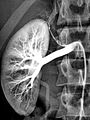
Arteriography of a healthy kidney


Airport security
Personal control
Devices for scanning people by means of X-rays are used for examinations in which objects such as weapons , explosives , smuggled goods or stolen goods such as drugs or diamonds are to be detected by the people. There are currently three types of X-ray scanner in use: backscatter systems , transmission systems and combined backscatter or transmission systems. With transmission systems, objects can be found that have been swallowed or are hidden in body cavities. As with medical applications, the X-ray contrast caused by the material-dependent X-ray opacity is evaluated. The radiopacity does not play a role in backscatter systems.
Baggage control

Baggage scanners are used for airport security. The picture on the right shows an X-ray image of a piece of luggage.
Materials science
While light, also electromagnetic radiation, only penetrates transparent substances, X-rays and gamma rays can penetrate non-transparent solid materials and provide information about hidden internal irregularities, cavities and inclusions.
astronomy
X-ray telescopes observe space in the range of X-rays. However, the interstellar gas absorbs radiation with wavelengths below 912 Å, the so-called Lyman's edge . This corresponds to the ionization energy of hydrogen in the ground state. Below about 100 Å , the interstellar medium begins to become transparent to electromagnetic radiation. While the absorption of helium and the K and I absorption edges of the more common elements are still hindered in the area of soft X-rays, below about 10 Å the interstellar medium is practically completely transparent. This means that interstellar sources (in contrast to the sun) cannot be observed in this area.
Individual evidence
- ↑ Manfred von Ardenne: electron microscopy physics · technology · results . Springer-Verlag, 2013, ISBN 978-3-642-47348-7 , pp. 127 ( limited preview in Google Book search).
- ↑ a b Detlef Kamke, Wilhelm Walcher: Physics for medical professionals . Springer-Verlag, 2013, ISBN 978-3-322-80144-9 , pp. 620 ( limited preview in Google Book search).
- ^ W. Bothe, PP Ewald, F. Kirchner, H. Kulenkampff, EG Steinke, H. Geiger: X-ray radiation Exclusively X-ray optics Volume XXIII · Second part . Springer-Verlag, 2013, ISBN 978-3-642-99594-1 , p. 4 ( limited preview in Google Book search).
- ↑ Peter Gängler, Thomas Hoffmann, Brita Willershausen: Conservative Dentistry and Periodontology . 3rd edition, Georg Thieme, 2010, ISBN 978-3-13-154073-7 , p. 183 ff. ( Limited preview in Google book search).
- ↑ Rolf Nolden; Ernst Sauerwein: Dentistry: preventive dentistry, cariology, endodontology, periodontology, children's dentistry . S. 142 Georg Thieme, 1994, ISBN 3-13-455606-5 .
- ↑ Hendrik Meyer-Lückel, Kim Ekstrand, Sebastian Paris: Caries: Science and Clinical Practice . Georg Thieme, 2012. ISBN 978-3-13-169321-1 , pp. 97 ff. ( Limited preview in the Google book search).
- ↑ Bernd Michael Balletshofer: Heart and vessels. Georg Thieme Verlag, 2006, ISBN 3-13-141131-7 , p. 125 ff. ( Limited preview in the Google book search).
- ↑ Sebastian Bertl, illustration with millimeter waves for identity checks , dissertation 2009, Technical University of Munich, Chair for High Frequency Technology. Retrieved February 1, 2017.
- ↑ Karlheinz Schiebold: Non-destructive material testing - radiographic testing . Springer-Verlag, 2014, ISBN 978-3-662-44669-0 ( limited preview in Google book search).
- ↑ Peter Schneider: Introduction to Extragalactic Astronomy and Cosmology . Springer-Verlag, 2006, ISBN 978-3-540-30589-7 , pp. 32 ( limited preview in Google Book search).
- ↑ A. Unsöld, B. Baschek: The new cosmos . Springer-Verlag, 2013, ISBN 978-3-662-06533-4 , pp. 415 ( limited preview in Google Book search).