Ranvier lace ring
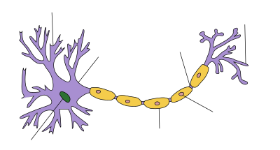
| Structure of a nerve cell |
|---|
The Ranvier-Schnürring [ rãviˈe ] - also called Ranvier'scher Schnürring or Ranvier-Node - is a section of a myelinated axon in which the cell membrane of the axon is exposed. A Ranvier ring can occur in the central nervous system between two myelin sections of an oligodendrocyte , or in the peripheral nervous system between two Schwann cells .
The Ranviersche Schnürring is named after the French anatomist Louis-Antoine Ranvier (1835-1922), who first presented it to the academy in 1871 ( Comptes rendus , 1871) and in March 1872 in his article Recherches sur l'histologie et la physiolige des nerfs described and illustrated in detail:
"Je me propose, dans ce mémoire, de décrire une disposition nouvelle des nerfs, que j'ai communiquée déjà, et de rechercher quelles sont, pour les tubes nerveux, les voies d'échange des matériaux de nutrition et de désassimilation"
.
construction
Ranvier rings have a length of approx. 1 μm and appear along the course of the axon at a distance of approx. 0.2–2 mm. The section between each two rings is called the internode , the section adjacent to the ring is called the paranodium .
Ranvier lacing rings are important for the rapid conduction of saltatory excitation . The action potential does not run continuously along the medullary nerve fiber , but “jumps” from ring to ring. The cell membrane in the area of the laced rings has a high density of voltage-controlled sodium channels and can generate a strong Na + influx during depolarization . Between these, the electrical excitation is transmitted electrotonically through the insulation of the medullary sheath . In the area of the paranodium, the axon membrane and myelin are firmly connected by binding proteins ( contactin , contactin associated protein).
The connection between the glial cell ( oligodendrocytes and astrocytes in the central nervous system (CNS), Schwann cells in the peripheral nervous system (PNS)) and the axon is closed on the sides of the constriction ring by para- nodal septate junctions . In this way, a small, closed space is created, the biochemical milieu of which can be well regulated and separated from the environment; Diffusion losses are minimized.
Web links
- Video: Electrophysiology of the Ranvier cord ring - isolation, assembly, morphology . Institute for Scientific Film (IWF) 1980, made available by the Technical Information Library (TIB), doi : 10.3203 / IWF / C-1413 .
Individual evidence
- ↑ Elior Peles, Sebastian Poliak: The local differentiation of myelinated axons at nodes of Ranvier . In: Nature Reviews Neuroscience . tape 4 , no. December 12 , 2003, ISSN 1471-0048 , p. 968–980 , doi : 10.1038 / nrn1253 ( nature.com [accessed May 7, 2019]).
- ↑ Matthew N. Rasband, Elior Peles: The Nodes of Ranvier: Molecular Assembly and Maintenance . In: Cold Spring Harbor Perspectives in Biology . tape 8 , no. 3 , September 9, 2015, ISSN 1943-0264 , doi : 10.1101 / cshperspect.a020495 , PMID 26354894 , PMC 4772103 (free full text) - ( cshlp.org [accessed May 7, 2019]).
- ↑ Research on l'histologie et la physiolige des nerfs. In: Archives des Physiologie Normale et Pathologique IV / 2 (Mars 1872), pp. 129–149
- ↑ Robert F. Schmidt: Outline of Neurophysiology . Springer-Verlag, 3rd edition 2013, ISBN 9783642962301 , p. 10.
- ↑ Gerhard Heldmaier, Gerhard Neuweiler: Comparative animal physiology: Neuro- and sensory physiology . Springer-Verlag, 2013, ISBN 9783642556999 , p. 46

