
Skeletal maturity
The skeletal maturity (also called skeletal age determination ) is a measure of the development of the adolescent. Normally the skeletal maturity to the chronological skeletal age also fits the age of the examinee, in the case of significant deviations is called developmental disorders such as precocious puberty , delayed puberty , gigantism and dwarfism .
There are several methods of determining maturity on a growing skeleton . What they have in common is the reference to a standard that was determined from a physically unrestricted, North American or British (white) population group.
In addition to the survey of the tooth status , the skeletal maturity is determined using an X-ray image . Determination using a hand exposure is the most common method, but determination using the knee joint is also described.
principle
The development of the hand skeleton usually follows a certain pattern (assuming normal development), with maturation taking place somewhat faster in girls than in boys.
In newborns there are no hand x-rays in the wrist of the girl, only the capitate and hamate are ossified (and thus visible) in boys , the other carpal bones are not yet ossified and only made of cartilaginous structure and therefore cannot be seen on the x-ray . No epiphyseal nuclei are visible in the area of the phalanges .
With advancing age and maturation, the structures mentioned above become increasingly ossified, usually in the following order in the wrist: capitate and hamate - triquetrum - lunate - trapezium - scaphoideum and trapezoideum . At the age of 14 (girls) or 16 (boys) years all growth plates of the hand are closed except for the epiphyseal plates of the distal radius and the ulna, i.e. the growth is largely complete.
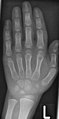
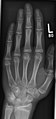
- X-rays of the hand bones of children of different ages

Boy, 3.5 years. Also note the brachymesophalangia on the little finger
Boy, 5 years
Girl, 14 years

Boy, 14 years


Methods

The two most common methods of determining skeletal age are the Greulich and Pyle methods and the Tanner and Whitehouse methods . With the atlas method according to Greulich and Pyle, the X-ray image of the left hand is compared with reference images from an atlas; the skeletal age is then derived from the image that comes closest to the current image. If the right hand is used, there is no significant difference in the determined skeletal age.
It is known from comparative studies that the skeletal age determinations using the two methods differ somewhat. The method according to Tanner and Whitehouse is considered more precise for German children, here a maturity score is created with the help of many reference images of individual sections of the left hand , from which the bone age can then be determined using a table. Determining the skeletal age according to Tanner and Whitehouse is therefore more complex.
Both methods can be determined automatically.
Due to the population on which the book atlases are based, the provisions in the context of statistical reliability only apply to these populations. The standards must be adapted for other populations.
An Israeli company offers an ultrasound system that measures the speed of sound conduction in the wrist, depending on the degree of ossification .
Indications
From a medical point of view, the correlation of skeletal maturity with chronological age is indicated in the case of disorders of development (accelerated or retarded) and growth as well as in certain other endocrinological diseases.
In addition to the medical indication, there is also a forensic indication . If juvenile offenders are caught who cannot (or do not want to) identify themselves, a decision must be made as to whether or not they can be arrested. If the age cannot be determined otherwise, an expert opinion on the skeletal age can be used. Due to the standard deviations (upwards and downwards usually at least one year), these reports are based on the minimum skeletal age (principle in dubio pro reo ), so that this is usually to the advantage of the accused.
Critical evaluation of forensic skeletal age determination and its application
A conclusion from the skeletal age to the chronological age is only possible to a very limited extent for the following reasons:
- The determined skeletal age is a statistical mean value (50th percentile ). It must be taken into account that there is a wide range of physiological fluctuations. ( Assuming a normal distribution , 68% of all patients lie within the single standard deviation and 95% within double the standard deviation.)
- The values of Greulich and Pyle were determined in the 1930s in upper and lower class children in Cleveland (USA). The values according to Tanner and Whitehouse in the 1950s in England, on children of the middle and lower classes.
- In cases of doubt, when assessing the need for help of refugees after an inspection of the teeth and external genital organs, the medical-radiological carpal bone examination is used to determine the age, which often leads to higher age assessments than stated by the refugee and thus the above-mentioned principle "In case of doubt for the Defendants "should contradict. Pro Asyl again writes: "If the age of a possibly minor cannot be determined with certainty, the principle of the protection of minors requires that the latest possible date of birth is assumed"
- According to an expert report commissioned by Pro Asyl and the Association of Democratic Doctors and Doctors, the informative value of the investigation is said to be "completely unsuitable for non-European young people" and "an inadmissible interference with the physical integrity of Art. 2 II 1 GG." (Göbel- Zimmermann, R. 1999: 143). However, with the amendment of the AuslG to the AufenthG together with the creation of §§ 49 III, VI, this inadmissible encroachment is no longer valid; instead, an X-ray examination to determine age is expressly permitted in VI, pp. 1 and 2 (Weichert, in: Huber, AufenthG, §49 Rn 25, 2010)
- The question must be asked: Do the standards also apply to the patient being examined? Play here z. B. besides the ethnic affiliation and the socioeconomic status of a role. There are comparative studies from Japan, Europe and the USA (on whites and colored people) which show that the skeletal age can vary by a good 1 year depending on ethnicity. While ethnic affiliation is now considered to be of secondary importance, it is known that a low socio-economic status leads to a development delay and thus to an underestimation of age. However, this usually does not result in any disadvantage for the person concerned.
- The values only apply to healthy children. For forensic reasons, however, the examination does not usually know whether the child is healthy or whether there are any factors that could have accelerated or slowed down skeletal maturation and this cannot be inferred from the X-ray image. In the case of certain diseases, it would be completely impossible to draw conclusions from the skeletal age. Even with long-term illnesses or malnutrition , skeletal development can lag significantly.
- In pediatric orthopedics, the X-ray of the hand is used as a meaningful diagnostic aid to determine the total body size, the completion of the growth in length and to determine deviations between skeletal and chronological age. In the spine, also known as is Risser sign common
However, it is more important to determine the period of the expected largest growth spurt in order to limit unnecessary and stressful therapeutic measures to the period that is absolutely necessary.
Delayed skeletal maturation
Possible causes of delayed skeletal maturation:
- Kleidocranial Dysplasia
- Hypothyroidism
- Kampomele dysplasia
- Spondylometaphyseal dysplasia
- Cerebral palsy
literature
- J. Christopher Bertozzi, Paul M. Bunch, Cree M. Gaskin, S. Lowell Kahn: Skeletal Development of the Hand and Wrist: A Radiographic Atlas and Digital Bone Age Companion. Oxford University Press, 2011, ISBN 978-0-19-978205-5 .
- JM Tanner , RH Whitehouse: Growth and Development Reference Charts (Tanner-Whitehouse Standards) . Castlemead Publications, 1984, ISBN 0-948555-00-9 .
- KC Grave, T. Brown: Skeletal Ossification and the Adolescent Growth Spurt . In: Am. J. Orthodont. No. 69 , 1976 (English).
- Andreas Ruhland: Orthodontic diagnostics . 2., completely redesigned. u. supplementary edition. Hanser, Munich / Vienna 1982, ISBN 3-446-13534-0 , p. 52 ff .
Individual evidence
- ↑ SI Pyle, NL Hoerr: Radiographic Atlas of Skeletal Development of the Knee. A Standard of Reference. Charles C Thomas Publ., 1955, OCLC 899039567 .
- ↑ DD Martin, J. Neuhof, OG Jenni, MB Ranke, HH Thodberg: Automatic Determination of Left- and Right-Hand Bone Age in the First Zurich Longitudinal Study. In: Hormone Research in Pediatrics. Vol. 74, 2010, pp. 50-55.
- ↑ HH Thodberg, S. Kreiborg, A. Juul, KD Pedersen: The BoneXpert Method for Automated Determination of Skeletal Maturity. In: IEEE Trans Medical Imaging. Vol. 28, 2009, pp. 52-66.
- ^ SY Zhang, G. Liu, CG Ma, YS Han, XZShen, RL Xu, HH Thodberg: Automated Determination of Bone Age in a Modern Chinese Population. In: ISRN Radiology. Volume 2013.
- ↑ HJ Mentzel, U. Unbehaun ao: Sonographic bone age determination in children and adolescents and comparison with various conventional methods. In: Fortschr Röntgenstr. 2008; 180 - VO_202_5 doi: 10.1055 / s-2008-1073446
- ↑ Pro asylum. 1995, p. 14. (library.fes.de)
- ^ Peter Kühne: On the situation of refugees in Germany . Bonn 2001, ISBN 3-86077-972-9 , (discussion group on migration and integration). FEuS Library, Electronic ed., 2002, fes.de (406 kB)
- ↑ F. Hefti: Pediatric Orthopedics in Practice . Springer, 1998, ISBN 3-540-61480-X , S, 648.