
Spondylolisthesis
| Classification according to ICD-10 | |
|---|---|
| M43.1 | Spondylolisthesis |
| ICD-10 online (WHO version 2019) | |
A spondylolisthesis (in German: spondylolisthesis ) or vertebral sliding is an instability of the spine in which the upper part of the spinal column with the sliding vertebra slides ventrally (forward) over the vertebral body below (ventrolisthesis or anterolisthesis) . In the opposite case, one speaks of a retrolisthesis .
The word is derived from the Greek : σπόνδυλος , "vortex" and ancient Greek ὀλίσθησις olisthesis , "glide". Therefore, the correct hyphenation is also Spondyl-Olisthesis, although Spondylo-Listhesis is also often used. Likewise, the short form should be "Olisthese" and not "Listhese".
The spondylolisthesis is often an incidental finding or only associated with minor discomfort. Depending on the strength of the vertebral sliding, single or multiple nerves in the spinal canal can be pinched and stretched in the long term . This can cause nerve damage and cause a nerve to malfunction. It can paralysis occur, both the legs and the function of the bladder and rectum concern. At the same time, the intervertebral disc ( herniated disc ) and vertebral joint ( spondylarthrosis ) in the corresponding segment wear out excessively, which can sometimes cause severe pain.
Differentiation according to cause and severity
causes
Spondylolisthesis can have various causes, so currently (2007) the following listed forms are known, of which in turn there are two subtypes:
- congenital forms (due to abnormal development or genetic makeup)
- dysplastic (malformed) form: This is a structural disorder of the lumbosacral (between the lumbar spine and sacrum ), which leads to the vertebral body slipping.
- Subtype: The dysplastic, axially aligned articular processes cannot prevent slipping.
- Subtype: The sagittally aligned vertebral joints enable ventral gliding.
- isthmic form: the interarticular portion (between the joints) of the vertebral arch is only cartilaginous - not ossified - and thus a weak point. A fracture ( lysis gap ) allows the vertebral body to slide off.
- Subtype: Repeatedly acting flexion - extension movements can cause the lysis gap and thus slipping.
- Subtype: A fracture that has healed once or several times and is caused by external impacts or loads with subsequent lengthening of the interarticular portion makes it possible for it to slip.
- acquired forms
- Degenerative form: Wear-related changes in the intervertebral space and / or vertebral joint cause the vertebral body to slide off.
- Traumatic form: an injury-related fracture outside the interarticular portion of the vertebral arch leads to the vertebral body sliding ventrally.
- Pathological form: A bone disease leads to reduced bone strength in the interarticular portion of the vertebral arch and, with a subsequent fracture, thus to the slipping of the vertebral body.
- Postoperative form: As a result of a spinal surgery, various changes in the operated segment can slide the vertebral body.
Degrees of severity
According to Meyerding (MD), there are four stages of difficulty:
- MD I °: offset of the vertebral bodies to one another by less than 25% of the vertebral body depth,
- MD II °: offset by 25-50%,
- MD III °: offset by 50-75%,
- MD IV °: offset by more than 75%.
If the vertebrae have lost contact with each other and the upper one slides freely forward and down, one speaks of a spondyloptosis (MD V °).
Diagnosis
Usually it is an incidental finding on the X-ray. In the case of back pain with radiating discomfort in the legs, an X-ray image can be made in two planes of the lumbar spine.
The position and posture of the person affected have an influence on the diagnosis. Under certain circumstances, vortex sliding can only occur with certain movements, so if there is any suspicion, it makes sense to perform functional recordings. These are two additional x-rays while standing in a forward position and in a reclined position.
The anatomy can be shown in detail with the CT or MRI , but the degree of severity can be underestimated. The MRI is particularly suitable for assessing the intervertebral discs and the nerves. In questionable cases, CT is particularly suitable to prove or exclude the bony defect (spondylolysis).

Image 1: Sliding between lumbar vertebrae 4/5, in the lateral x-ray.
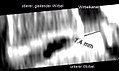
Image 2: Sliding vertebrae, stage 1, in the CT.
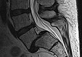
Image 3: Sliding vertebrae L5 / S1, stages 2–3, MR sagittal section, spinal canal free.

Image 4: The nerve exit hole L5 is narrowed.


Figure 1 is the conventional X-ray of a vertebral sliding between vertebrae 4 and 5 of the lumbar spine. The degree of severity lies on the border between stages 1 and 2. On the right in the picture, the bony interruption of the vertebral arch ( spondylolysis ) can also be seen. So it is a "correct" spondylolisthesis (also called spondylolisthesis vera).
Figure 2 shows the sagittal reconstruction of an almost horizontal CT examination of the lower lumbar spine. The upper vertebral body has slipped 7.4 mm from the lower one, the intervertebral disc tissue is severely degenerated, which is why it sometimes appears as a black spot (vacuum phenomenon). The spinal canal is narrowed, the dural sac is narrowed here . The upper vertebra slides downwards with the intervertebral disc.
Figure 3 shows L5 / S1 vertebral sliding laterally in the MR . The vertebrae are offset by 18 mm (stages 2-3). The intervertebral disc is deformed, the height of the intervertebral disc space L5 / S1 is reduced. The spinal canal is dilated. This is typical of a "real" spondylolisthesis.
Figure 4 is from the same investigation as Figure 3. It shows a foramen - stenosis that can stand with the spondylolisthesis related. The spinal nerve L5 (yellow circle) is thereby raised ; this can explain severe complaints in the supply area of this nerve.
treatment
There are currently no valid guidelines for the treatment of spondylolisthesis. The procedure is based on the study situation or the personal experience of the attending physician. Back exercises that strengthen the back muscles and reduce lordosis are common .
It is essential for the therapy decision to establish whether it is a real spondylolisthesis with spondylolysis or a pseudospondylolisthesis z. B. acts with accompanying spinal stenosis .
Non-operative therapy
Because of the unclear state of the study, surgery should initially not be used.
In addition to pain medication, pain treatment can also be carried out using infiltration therapy or PRT . Medical massages can also help relieve pain. The administration of muscle relaxants has no positive effect.
In some cases, a trunk orthotic is suitable to alleviate the symptoms. Stabilizing the muscles through physiotherapy can be helpful.
Operative treatment

Surgical therapy can be considered, though
- the pain cannot be managed conservatively,
- the vortex slip increases rapidly in a short time,
- muscular failures occur or
- urinary retention or fecal incontinence occurs.
The surgeon tries during the operation z. B. to return the vertebral body to its original position and then to block it ( spinal fusion ). This major procedure is usually performed through an anterior ( ventral ) or posterior ( dorsal ) spinal access and in one or two sessions. Whether an operation is really necessary and which procedure is used depends on the symptoms and is determined by the attending physician.
In addition to the basic surgical risks, there is also the risk of nerve damage from the screws inserted and the risk of postoperative scarring, which may cause more pain than the underlying disease. These complications are summarized as failed back surgery syndromes.
literature
- JE Buck: Direct repair of the defect in spondylolisthesis . In: JBJS . [Br] 52-B, pp. 432-437 (1970).
- SPF Hughes , R. Döhler , KM Tan, HJ Watson, JHS Scott: Lateral mass fusion for lower back pain (Wiltse, spondylolisthese). In: Archives of Orthopedic and Traumatic Surgery . 106, pp. 381-384 (1987).
- Fritz Hefti: Pediatric Orthopedics in Practice . Springer, 1998, ISBN 3-540-61480-X .
- Adam Greenspan: Orthopedic Radiology. A practical approach. 3. Edition. Lippincott Williams & Wilkins, 2000, ISBN 0-7817-1589-X .