Aglaophyton major
| Aglaophyton major | ||||||||||||
|---|---|---|---|---|---|---|---|---|---|---|---|---|

Aglaophyton major , reconstruction |
||||||||||||
| Temporal occurrence | ||||||||||||
| Devon , Emsium / Eifelium | ||||||||||||
| approx. 396 ± 8 million years | ||||||||||||
| Locations | ||||||||||||
| Systematics | ||||||||||||
|
||||||||||||
| Scientific name of the genus | ||||||||||||
| Aglaophyton | ||||||||||||
| DS Edwards | ||||||||||||
| Scientific name of the species | ||||||||||||
| Aglaophyton major | ||||||||||||
| DS Edwards |
Aglaophyton major is a fossil plant species that, due to its combination of characteristics, stands between mosses and vascular plants . It wasfirst describedin 1920 from the Rhynie Chert in the Scottish county of Aberdeenshire . She alone forms the genus Aglaophyton .
features
Sporophyte
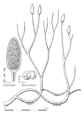
The rhizome grows horizontally. Rhizoids that are not septate and that arise from epidermal cells grow in certain places .
The shoots of the sporophyte are upright, naked and branch evenly dichotomously. The plant is a maximum of 18 centimeters high. The diameter of the rung is around six millimeters.
The conductive tissue consists of an inner zone of elongated, thin-walled cells and an outer zone of elongated, thick-walled cells. This water conduction tissue does not contain any tracheids because the cells lack the corresponding thickening. This central part is surrounded by a six-cell-thick layer of thin-walled, elongated cells, which are regarded as a " phloem " due to their position . Since this conductive tissue lacks the typical conductive elements (such as tracheids), they are also referred to as hydroids and leptoids, as is the case with mosses . The cortex connects to the outside and has extremely thick-walled cells in the outer area. The epidermis is made up of elongated cells. The stomata are formed by two companion cells, which are surrounded by six to eight slightly modified epidermal cells.
The sporangia are spindle-shaped and radially symmetrical in cross-section. They are terminal, i.e. at the tips of the axes, just above a dichotomous branching of the axis. The wall of the sporangia consists of three cell layers, so it is eusporangiat. The outer layer consists of thin-walled epidermal cells. This also has stomata. The next layer is parenchymal and similar to the inner cortex of the axis. The third layer surrounds the central spore mass as a tapetum layer. There are no pre-formed openings in the sporangium. The spores are 52 to 78 micrometers in size, trilet (have a three-pointed scar) and laevigat (smooth).
Gametophyte
In 1980 Renate Remy and Winfried Remy described Lyonophyton rhyniensis , which is probably the gametophyte of Aglaophyton . Only the distal end of the antheridia carrier is known. It is a bare shaft at least 16 mm long that ends in a disk-shaped structure 2.6 to 9 mm in diameter. The rounded antheridia sit on their surface. Each antheridium has a central, sterile tissue. The spermatozoids are often visible in the antheridia .
The gametophyte was independent, i.e. not connected to the sporophyte. The connection to the sporophyte is established by the similarities of the cuticle , the stomata and the anatomy of the water-conducting cells.
The female gametophyte has also been identified, but not yet published.
ecology
Aglaophyton grew singly or in groups on dry substrates covered with litter. Humid conditions were likely required for germination.
Systematic position
Aglaophyton is not a vascular plant . It corresponds to the structure of vascular plants with the exception of the structure of the conductive tissue. It thus occupies a middle position between mosses and vascular plants. Aglaophyton is traditionally placed with the Rhyniophyta, but these are a polyphyletic group and actually part of the vascular plants. In cladistic analyzes, Aglaiophyton is the sister group of vascular plants.
Botanical history
The fossils from the Rhynie Chert were initially mistaken for Rhynia gwynne-vaughanii , and in 1920 Robert Kidston and WH Lang described them as Rhynia major . For a long time no sporangia were known from Aglaophyton , which led to the assumption that Rhynia major was the gametophyte of Rhynia gwynne-vaughanii . This would have been a confirmation of a theory about the origin of land plants, which put similarly built (isomorphic) gameto- and sporophytes at the origin. The discovery of Rhynia major fossils with sporangia made further discussion unnecessary.
In 1986, Dianne S. Edwards was able to show on the basis of a new examination of the original material and on the basis of new discoveries that Rhynia major clearly did not have vascular bundles. She established the new, monotypical genus Aglaophyton .
supporting documents
- Paul Kenrick, Peter R. Crane: The Origin and Early Diversification of Land Plants. A Cladistic Study . Smithsonian Institution Press, Washington DC 1997, pp. 320-322. ISBN 1-56098-729-4
- Thomas N. Taylor, Edith L. Taylor: The Biology and Evolution of Fossil Plants . Prentice Hall, Englewood Cliffs 1993, pp. 196-198, 227. ISBN 0-13-651589-4
Individual evidence
- ↑ a b Aglaophyton on the Rhynie page of the University of Aberdeen
- ↑ Paul Selden , John Nudds: Window on Evolution. Famous fossil sites in the world . Elsevier Spektrum Akademischer Verlag, Munich 2007, p. 52. ISBN 978-3-8274-1771-8
- ↑ Kenrick, Crane: The Origin and Early Diversification of Land Plants , 1997, Fig. 4.31; Peter R. Crane, Patrick Herendeen, Else Marie Friis: Fossils and Plant Phylogeny . In: American Journal of Botany , Volume 91 (10), 2004, pp. 1683-1699.
- ^ DS Edwards: Aglaophyton major, a non-vascular land-plant from the Devonian Rhynie chert . In: Botanical Journal of the Linnean Society . No. 93 , 1986, pp. 173-204 .