Babesia
| Babesia | ||||||||||||
|---|---|---|---|---|---|---|---|---|---|---|---|---|

Babesia ( Babesia ) |
||||||||||||
| Systematics | ||||||||||||
|
||||||||||||
| Scientific name | ||||||||||||
| Babesia | ||||||||||||
| Starcovici , 1893 |
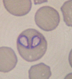
Babesia ( Babesia ) belong to the unicellular Apicomplexa . They are pathogens and parasitize on the red blood cells of vertebrates . Babesia is transmitted by different types of tick and causes babesiosis in humans and animals . They are named after their discoverer, Victor Babes .
Propagation cycle
Babesia shows an obligatory change of host. The asexual phase of reproduction ( merogony ) takes place in the erythrocytes (red blood cells) of the vertebrates, sexual reproduction ( gamogony ) and then several asexual divisions ( sporogony ) in the tick.
After the sporozoites have been transferred to the vertebrate via the tick's saliva, they penetrate the erythrocytes. Here they develop into trophozoites . These are divided by DNA - replication to binuclear stages of development, and finally by cell division to merozoites (merogony). After the erythrocytes have been destroyed, the merozoites are released and can then penetrate into new, as yet unaffected erythrocytes. However, some stages of the parasite do not divide in the erythrocytes, but rather form into so-called gamonts .
Ticks that suckle on infected animals take the babesia back with the erythrocytes. In the tick's intestinal sac, the babesia is released from the erythrocytes. In the tick's intestine, they multiply with multiple nucleus division and the formation of large division forms ( gamonts ), which divide into mononuclear gametes . These gametes fuse with another to form a zygote (gamogony). The zygotes penetrate the intestinal epithelial cells of the ticks and develop into mobile kinetes , which via the hemolymph attack all organs of the tick, including the salivary glands and ovaries. Further asexual division steps take place in the organs, through which the sporokinets arise. In the tick's salivary glands, these divide into thousands of sporozoites, which are transferred to a new host with the next blood meal.
Systematics
A distinction is made between “small” (<3 µm) and “large” (> 3 µm) Babesia.
| Art | author | host | Occurrence |
|---|---|---|---|
| Babesia bigemina | (Smith & Kilborne 1893) | Cattle (" Texas fever "), zebu , buffalo , red deer | Southern Europe, America, Asia, Africa |
| Babesia bovis | (Babès 1888) | Cattle, zebu, red deer, roe deer | Southern Europe, America, Asia, Africa |
| Babesia canis | (Piana & Galli-Valerio 1895) | dogs | worldwide ( babesiosis of the dog ) |
| Babesia capreoli | Enigk & Friedhoff 1962 | Roe deer | ? |
| Babesia divergens | (M'Fadyean & Stockman 1911) | Cattle (“Mairot” or “Weiderot”), roe deer, fallow deer and red deer, primates , humans | Europe, North Africa |
| Babesia duncani | (Conrad et al. 2006) | human | North America |
| Babesia equi | Laveran 1901 | Horses | ? |
| Babesia gibsoni | (Patton 1910) | dog | Asia, USA ( babesiosis of the dog ) |
| Babesia jakimovi | (Nikolskii et al. 1977) | Beef | Siberia |
| Babesia major | (Sergent et al. 1926) | Beef | Europe, Asia |
| Babesia motasi | (Weynon 1926) | Sheep , goats | ? |
| Babesia occultans | Gray & de Vos 1981 | Beef | South Africa |
| Babesia ovis | (Babès 1888) | Sheep, goats | ? |
| Babesia ovata | Minami & Ishihara 1980 | Beef | East asia |
| Babesia vulpes ( Theileria annae ) | Baneth et al. 2015 | Fox | North America, Southern Europe |
The species B. microti , which used to belong to the Babesia , is now counted as Theileria microti to the Theileries .
Diseases (babesiosis)
Babesia can be transmitted by ticks, but also by blood transfusions. The destruction (bursting) of the erythrocytes leads to the symptoms characteristic of babesiosis : fever , anemia ( anemia ) and jaundice (jaundice). In (per) acute cases, urine is often coffee-brown to red in color as a symptom of the disease ( hemoglobinuria , not hematuria).
literature
- Vogl, S .: Molecular-phylogenetic differentiation of babesia of cattle. Dissertation of the Veterinary Faculty, Ludwig Maximilians University Munich , 2004. ( pdf )
Individual evidence
- ↑ Gad Baneth, corresponding author Monica Florin-Christensen, Luís Cardoso, and Leonhard Schnittger: Reclassification of Theileria annae as Babesia vulpes sp. nov. In: Parasite Vectors. 2015; 8: 207 PMC 4393874 (free full text).
- ↑ BL Herwaldt, DF Neitzel, JB Gorlin, KA Jensen, EH Perry, WR Peglow, SB Slemenda, KY Won, EK Nace, NJ Pieniazek, M. Wilson: Transmission of Babesia microti in Minnesota through four blood donations from the same donor over a 6-month period. In: transfusion . Volume 42, Number 9, September 2002, pp. 1154-1158, ISSN 0041-1132 . PMID 12430672 .