Escherichia virus lambda
| Escherichia virus lambda | ||||||||||||||||||
|---|---|---|---|---|---|---|---|---|---|---|---|---|---|---|---|---|---|---|
| Systematics | ||||||||||||||||||
|
||||||||||||||||||
| Taxonomic characteristics | ||||||||||||||||||
|
||||||||||||||||||
| Scientific name | ||||||||||||||||||
| Escherichia virus Lambda | ||||||||||||||||||
| Short name | ||||||||||||||||||
| λ | ||||||||||||||||||
| Left | ||||||||||||||||||
|
The Escherichia virus lambda (English Escherichia phage lambda , formerly also known as lambda phage , bacteriophage lambda , Enterobacteria phage lambda or phage λ ) is a virus from the order Caudovirales that is hosted by the bacterium Escherichia coli ; and is therefore classified as bacteriophage . It is of great importance in the history of virus research and molecular genetics as well as in genetic engineering . It was discovered by Esther Lederberg in 1950 .
morphology
The virion of Escherichia virus Lambda is as with all Siphoviridae of a capsid constructed with the dsDNA containing and a non-contractile tail portion. The capsid has isometric, icosahedral symmetry and is about 60 nm in diameter; it consists of 72 capsomers (60 hexamers, 12 pentamers, triangulation number T = 7). Due to the gap for the attachment of the tail part, the capsid does not consist of 420 (= 60 × 6 + 12 × 5) molecules of the two main capsid proteins E and D, but only of 415.
The tail part is flexible but non-contractile and 8 × 150 nm in size. At its end there are four long fibers, which however disappear in strains grown in vitro .
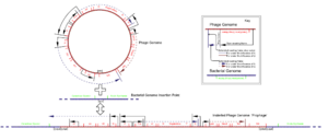
The linear, double-stranded DNA genome of the lambda phage is 48,502 bp in size and codes for about 70 proteins. The complete genome sequence was determined by Frederick Sanger in 1982. The linear DNA has short single-stranded sections at both ends, which are complementary to one another and serve to recirculate the phage DNA after injection into the host. These ends are also called cohesive ends (cos), homologous binding ends or sticky ends (engl. Sticky ends ), respectively.
integration
The Escherichia virus lambda is a temperate bacteriophage . After infection of the host cell, its linear phage DNA is closed into a circular DNA by joining the sticky ends of 12 bases each at the cos site of the DNA by the bacterium's DNA ligase . The circular DNA can be incorporated into the bacterial chromosome by the viral integrase . The phage DNA is integrated into the host genome via the att site of the plasmid and is a site-specific recombination and can lead to specialized transduction . In this state, the lambda phage is a prophage and remains integrated into the host genome without expressing its genes .
Expression

The expression of the prophage genes is suppressed by a repressor protein. The only gene that remains active is the repressor gene cI itself. This means that the prophage DNA is also duplicated every time the host cell divides, but its genes are not transcribed . This process is also known as the lysogenic cycle . The termination of this inactive lysogeny can be caused by stress factors such as B. antibiotics, UV radiation, nutrient deficiencies and especially DNA damage can be induced. In the latter case, damage to the genetic information leads to the activation of RecA, an inducer of DNA repair processes, the SOS response . However, after activation, RecA not only cleaves its actual substrate, but also the lambda repressor cI. This can no longer dimerize, which leads to the termination of the repression and thus also to the expression of the phage genome. The lambda phage can now enter the lytic cycle , during which the actual reproduction of the phage takes place. After the phage components have been synthesized and assembled, the cells are lysed and thus the mature, infectious phages are released.
Applications in genetic engineering
Lambda phages are used as insertion vectors in genetic engineering . However, some modifications are necessary before using it as a vector. First of all, excess restriction sites should be removed. In order to insert an additional gene segment (insert), the original phage sequences must be removed, since otherwise the capsid cannot be packaged due to the artificial genome being too large. For this purpose, phage genes that are not required for replication and the genes for the lysogenic cycle are removed. This creates space for an approx. 8kb insert. Escherichia virus lambda is used both as a cloning vector for the production of cDNA libraries and as a platform for phage display .
The isolated DNA of the Escherichia virus lambda is also used as a size marker for agarose gel electrophoresis . The digestion with a restriction endonuclease results in a mixture of DNA fragments of different sizes of known size, which allow the size of other DNA fragments to be determined after their separation. The restriction enzyme PstI is often used here, since the fragment mixture produced in this way covers a relatively wide range of sizes.
literature
- Court, DL et al. (2007): A new look at bacteriophage lambda genetic networks. In: J. Bacteriol. Vol. 189, 298-304. PMID 17085553 doi : 10.1128 / JB.01215-06 PDF
- Gottesman, ME & Weisberg, RA (2004): Little lambda, who made thee? In: Microbiol. Mol. Biol. Rev. Vol. 68, pp. 796-813. PMID 15590784 doi : 10.1128 / MMBR.68.4.796-813.2004 PDF
- Coleclough, C. & Erlitz, FL (1985): Use of primer-restriction-end adapters in a novel cDNA cloning strategy. In: Genes. Vol. 34, pp. 305-314. PMID 2408965
- Hohn, B. (1983): DNA sequences necessary for packaging of bacteriophage lambda DNA. In: Proc. Natl. Acad. Sci. USA Vol. 80, pp. 7456-7460. PMID 6324174 , PMC 389970 (free full text)
- Sanger, F., Coulson, AR, Hong, GF, Hill, DF, Petersen GB (1982): Nucleotide sequence of bacteriophage lambda DNA. In: J. Mol. Biol. Vol. 162, pp. 729-773. PMID 6221115
Web links
Individual evidence
- ↑ a b c d ICTV: ICTV Taxonomy history: Enterobacteria phage T4 , EC 51, Berlin, Germany, July 2019; Email ratification March 2020 (MSL # 35)
- ↑ Invisible Esther jax.org
- ^ Lambda phage. Retrieved January 15, 2020 .

