
Bacteriophage

As bacteriophages or simply phages ( singular phage , the ; of ancient Greek βακτήριον baktērion , Chopsticks 'and φαγεῖν phagein , eat') refers to various groups of viruses that on bacteria as host cells are specialized. According to the host specificity, the phages are divided into taxonomic groups, for example coli , staphylococci , diphtheria or Salmonella bacteriophages. With an estimated number of 10 30 virions in all seawater, phages are more common than any type of living being and form what is known as virioplankton .
Please note: Viruses are not living beings because they do not have their own metabolism . They are called "close to life" by some scientists.
history
The effect of phages was first described in 1917 by the Canadian Félix Hubert d'Hérelle . Although the Englishman Frederick Twort had already observed decomposition processes in staphylococcal cultures in 1915 , which can be attributed to the action of bacteriophages, his publication was practically ignored. D'Hérelle is therefore one of the discoverers of bacteriophages, the so-called "bacteria-eaters", alongside Frederick Twort. However, they owe their name and their discovery to d'Hérelle. In parallel to d'Hérelle, the German microbiologist Philalethes Kuhn postulated the existence of bacterial parasites based on observations of changes in bacterial cultures under certain conditions. He referred to this as Pettenkoferien and saw the "invisible microbe that counteracts the dysentery bacillus" described by d'Hérelle as a special case of these parasites. As it turned out later, his observations were not based on the existence of a bacterial parasite, but only on changes in the shape of the bacteria he was studying.
D'Hérelle imagined the bacteriophage to be an "ultravisible, corpuscular living being" that exists in a basic form and that adapts to different hosts, i.e. bacteria. In fact, as far as we know today, bacteriophages are highly specialized viruses that are bound to a specific host. Even if we are talking about hosts in this context, according to today's definition, bacteriophages, as viruses are not living beings, are not parasites . The first phages to be examined were seven phages from the bacterium Escherichia coli . They were named in the order of their discovery as Type 1 (T1), Type 2 (T2), and so on.
construction

The shape of the bacteriophages was mainly elucidated on the phages of the T series (T series) of Escherichia coli . The Coliphage T2 consists of a polyhedral head 100 nm in length with a tail of approximately the same length. Bacteriophages are grouped taxonomically according to their morphology , genome, and host . A distinction is made between DNA phages with single-stranded DNA, so-called ss-DNA phages (from English single-stranded ), and those with double-stranded DNA, so-called ds-DNA phages (from English double-stranded ). The Escherichia coli phages of the T series treated here as an example belong to the latter group.
The so-called T phages (e.g. T4 phage ) are distinguished from other bacteriophages by a relatively complex structure. Basically, they consist of a base plate (9), an injection device (injection device, 2) and a head (1) consisting of the so-called capsid (4) and the nucleic acid (3) contained therein. The head and injection device modules are connected by a neck (collar, 5). The base plate (which, like the capsid and injection device, is made up of proteins ) is covered with tail fibers (7) and spikes (8), which are used for adsorption on the host cell wall . The injection device consists of a thin tube, also called a tail tube (6), through which the phage DNA (3) is injected into the host cell. The tube is encased in a contractile tail sheath that contracts during injection. The capsid is composed of 152 capsomeres with icosahedral symmetry and contains the DNA of the phage. Due to this structure, the phages of the genus T4-like viruses (family Myoviridae ) are among the structurally most complex viruses.

Phages with single-stranded DNA, on the other hand, are usually small, spherical and tailless or filamentous. The RNA phages that also occur usually consist (if described up to this point in time) of a protein envelope that encloses a single-stranded RNA molecule. The diameter of these phages is around 25 nm, making them one of the smallest phages.

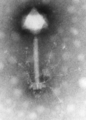
E. coli T2 bacteriophage ; Axially cut capsid

Lysehof of Bacillus phage gamma in Bacillus anthracis , on the right uninfected single colony
Synechococcus phage S-PM2 from seawater
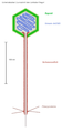
Lambda phage ( Escherichia virus lambda ); a schematic cross section


Multiplication

In the absence of a metabolism of their own, viruses require a host for reproduction , in the case of bacteriophages, a suitable, living bacterial cell. The reproduction can be divided into five phases:
- Adsorption on specific cell wall receptors : During adsorption, the ends of the tail fibers couple to suitable molecules ( receptors ) on the surface of the bacterium.
- Injection of the phage nucleic acid into the host cell: The phage's own nucleic acid , DNA or RNA , gets into the bacterium. The now functionless proteins of the empty phage envelope remain outside on the surface of the bacterium.
- Latency phase: During this phase, no phages can be detected in the bacterium. Now the transcription of the virus genome , the translation of the viral mRNA and the replication of the virus nucleic acid begin. This process takes a few hours at most.
- Production phase : After the phage genes have become active in a fixed order, all virus components, envelope proteins and tail fibers are formed.
- Maturity phase: In this phase of morphogenesis , the assembly takes place to form mature phage particles. First a head part, the capsid , is formed. The proteins inside serve as placeholders and are later replaced by the phage nucleic acid that penetrates the capsid. The nucleic acid threads take on a space-saving shape like a ball of wool.
- Release: The finished virus particles are released by enzymatic dissolution of the host cell. The lysozyme , which was formed by the reprogrammed bacterium, dissolves the bacterial murine cell wall . The cell bursts and about 200 infectious phage are released.
In the case of some phage types, the reproduction does not always proceed according to the lytic scheme described above . In the case of temperate phages, a distinction is made between lysogenic and lytic growth cycles or infection cycles. In a lysogenic cycle, the phage DNA is built into the bacterium's chromosome , creating a prophage . With each subsequent cell division, the genes of the phage and those of the bacterium are duplicated and passed on together. This cycle can later lead to the lytic cycle.
Giant phages
Double-stranded DNA phages with a genome size of more than 540 kbp are called megaphages, smaller ones with more than 200 kbp are called jumbo phages. In 2018/2019, the authors examined the faeces of people in Bangladesh and Tanzania , as well as baboons in Africa and pigs in Denmark. The samples contained bacteria of the genus Prevotella ( Prevotellaceae ), which were infected by a number of dsDNA megaphages, which the authors named “ Lak phages ” (after the location of Laksam Upazila , Bangladesh). The phages found were designated (provisionally) as Lak-A1, Lak-A2, Lak-B1 to Lak-B9 and Lak-C1. There could be a loose phylogenetic relationship to " Sphingomonas Phage PAU " (this giant phage infects bacteria of the species Sphingomonas paucimobilis , Sphingomonadaceae ) and thus to the phage family Myoviridae . The authors come to the conclusion that " lac phages " are "widespread, but previously overlooked members of the gut microbiome".
In February 2020, Basem Al-Shayeb and colleagues published an analysis that continues these investigations. In it they draw the limit for megaphages at 500 kb (which obviously means base pairs in the double-stranded case and bases or nucleotides in the single-stranded case). The authors prefer to consider all phages with more than 200 kb (ie jumbo phages and megaphages) as " English huge phages " (translated here as giant phages ). The authors identified a series of ten clades among this group , for which they suggested the following names: " Kabirphage ", " Mahaphage " (including the group of the " Lak phages "), " Biggiephage ", " Dakhmphage ", " Kyodaiphage ", " Kaempephage ", " Jabbarphage ", " Enormephage ", " Judaphage " and " Whopperphage " (all names refer to "huge" or " huge " in the different languages of the authors). Through their metagenomic analyzes of various samples, they were able to identify 351 dsDNA phage sequences, of which only 3 were less than 200 kb. The largest genome was 735 kb in length (a Mahaphage, which is apparently a new record; the previous one was 596 kb); ordinary non-giant phages have an average of only 52 kb. Some giant phages seem to use a non-standard genetic code in which the stop codon UAG codes for an amino acid . The hosts are (mostly) bacteria of the Firmicutes or the Proteobacteria , but also - as with the members of the Mahaphage group with the “ Lak phages ” - the Bacteroidetes . The genome codes for tRNAs in addition to the proteins commonly used in phage . The phages also interact in the CRISPR / Cas system (see CRISPR , CRISPR / Cas method , genome editing ): all major types of the system were represented, but most of the phages appeared to use the host's Cas proteins to act on themselves protect. In addition, the phages appeared to aid the hosts' CRISPR immune system in fighting off competing phages. Some Pseudomonas -infecting phages also code for anti-CRISPRs (Acrs) and proteins that form a nucleus-like compartment in which the phage can replicate its genome more independently of the host (see Viroplasma ). The authors see their work as further evidence of the worldwide distribution of the giant phages. They found evidence that the phages migrated between different hosts and ecosystems, which has implications for the spread of toxin and antibiotic resistance genes. Your CRISPR tools could be used in the future to improve the "gene scissors" CRISPR / Cas and to expand their functionality.
Tailless phage
For a long time, research only looked at members of the order Caudovirales , whose representatives are phages (bacterial and archaeal viruses) with a head-to-tail structure. Only recently have "tailless" phages been the subject of research. Some representatives are:
- Family Finnlakeviridae (ssDNA, with species Flavobacterium virus FLiP alias Phage FLiP )
- Family " Autolykiviridae " (dsDNA, suggested)
Haloviruses
Phages that attack halophilic bacteria and archaea are classified under the informal (non-taxonomic) term haloviruses ( English haloviruses ) . In addition to the genus Myohalovirus ( Caudovirales : Myoviridae ) with the species Halobacterium virus phiH confirmed by the ICTV and the proposed species " Halorubrum phage HF2 ", these are further unclassified and also as yet unconfirmed species "HF1", "HCTV-1", "2" and "5", "HGTV-1", "HHTV-1" and "2", "HRTV-4", "5", "7" and "8" ( caudovirales ), "HSTV-1" ( caudovirales : Podoviridae ) and "2" ( Caudovirales : Myoviridae ), "HVTV-1" ( Caudovirales : Siphoviridae ), " Halovirus VNH-1 " ("VNH-1", Fuselloviridae ) and " Haloferax tailed virus 1 " (HFTV1, Caudovirales ) .
Magroviruses
Marine archaea of the Euryarchaeota are classified as Marine Group ( English Marine Group ) II (MG-II, consisting of MG-IIa to MG-IId), III (MG-III) and IV (MG-IV) - the Marine Group I ( MG-I) denotes marine archaea of the Thaumarchaeota .
The non-taxonomic designation Magroviruses ( English magroviruses , MArine GROUP II viruses ) is used to classify phages that parasitize Euryarchaeota of the first group mentioned MG-II. These are dsDNA viruses with a genome size of 65-100 kbp with a head-to-tail structure: " Magrovirus A ", " Magrovirus B1 " and " B2 ", as well as " Magrovirus C " and (suspected) " Magrovirus D ".
application areas
Phages have found a wide range of applications in medicine , biology , agricultural sciences , especially in the area of genetic engineering . For example, phages are used in medicine to identify bacterial pathogens because of their host specificity . This procedure is called a lysotype . Due to the increasing frequency of multiple antibiotic resistances , intensive research is currently being carried out on the use of bacteriophages as antibiotic substitutes in human medicine (see: phage therapy ). Problems arise here from the poor stability of phages in the body, since they are eliminated as foreign bodies by phagocytes in a very short time . Felix d'Hérelle (see above) discovered this application of phages for the treatment of bacterial infections long before the discovery of penicillin and antibiotics . Later, however, with the introduction of chemotherapy using antibiotics, phage therapy was considered impractical and was forgotten. D'Hérelle founded the Eliava Institute for Phage Research in Georgia in 1934 together with the Georgian microbiologist Georgi Eliava , which still exists today. Today, phage therapy for otherwise therapy-resistant bacterial infections is carried out there and at the Ludwik Hirszfeld Institute for Immunology and Experimental Therapy in Wroclaw (part of the Polish Academy of Sciences ) . In Germany, the use for therapeutic purposes is not yet permitted.
The applications in food production are diverse; For example, a phage spray is used when packing sausages or slicing cheese.
In genetic engineering , temperate phages are used as vectors (e.g. phage λ ). For this purpose, phages are prepared in such a way that the genes that cause virulence are taken from their genome and replaced by genes that are of interest for genetic engineering, such as genes that are required for insulin production. These modified phages are then brought into contact with suitable bacteria, for example E. coli . After checking whether the desired gene has been integrated into the genetic material of the bacterial genome (gene-expressed antibiotic resistances are used, which are connected to the desired genes to be cloned ), the modified bacterial cells can be further cultivated and the insulin produced in this case can be isolated. Similarly, phages are used in agricultural technology to transduce certain genes in crops . An important application in biochemistry is the phage display for the identification of binding partners, e.g. B. in the isolation of new active ingredients.
However, the transformation of free DNA, which nowadays is mainly used for transfer into bacterial cells, is easier than using phages .
Phages and components are used for the removal of microbial contamination in food (e.g. by affinity magnetic separation ) as well as laboratory samples contaminated with endotoxins . Furthermore, there are human diagnostic applications, especially in the clinical area for the decolonization of pathogenic hospital germs such as MRSA . The phage proteins can be optimized for the respective application by means of protein design.
Possible economic damage
Bacteriophages can cause damage wherever bacterial processes serve humans and are desirable. Infection of lactic acid bacteria (LAB) by phages from raw milk is the most common cause of reduced or absent enzyme activity in starter cultures for cheese or curd milk production .
classification
The prokarytic viruses (bacterial and archaeal viruses, "bacteriophages") do not form a closed family group ( taxon ).
Baltimore classification
According to the Baltimore classification , phages can be grouped as follows:
- dsDNA bacteriophages:
- Family: Myoviridae
- Family: Siphoviridae
- Family: Podoviridae
- Family: Tectiviridae
- Family: Corticoviridae
- Family: Plasmaviridae
- Family: Lipothrixviridae
- Family: Rudiviridae
- Family: Fuselloviridae
- Family: Halspiviridae (with genus: Salterprovirus and species Salterprovirus His1 alias His 1 virus )
- Family: Guttaviridae
- Family: Akkermanviridae
- Family: Bicaudaviridae
- Family Thaspiviridae (with genus Nitmarvirus and species Nitmarvirus NSV1 alias Nitrosopumilus maritimus virus 1 )
- Family " Autolykiviridae " (suggested, see above)
- ssDNA bacteriophages:
- Family Finnlakeviridae
- Family: Inoviridae
- Family: Microviridae
- Special case
- Family Pleolipoviridae (with genus Gammapleolipovirus and species Gammapleolipovirus His2 alias His 2 virus , Haloarcula virus His2 )
- dsRNA bacteriophages:
- Family: Cystoviridae
- ssRNA bacteriophages:
- Family: Leviviridae
Taxonomic classification according to ICTV
In the system of virus taxonomy according to the International Committee on Taxonomy of Viruses (ICTV) , phages can be found in the following taxonomic groups:
| Area | order | family | morphology | Genome | Examples |
|---|---|---|---|---|---|
| Riboviria | Levivirales | Leviviridae | un enveloped , isometric | ssRNA , linear | MS2 , Qβ |
| Mindivirales | Cystoviridae | enveloped, spherical | dsRNA , segmented | Phi6 | |
| Varidnaviria | Belfryvirales | Turriviridae | wrapped, isometric | dsDNA , linear | STIV1 |
| Halopanivirals | Sphaerolipoviridae | wrapped, isometric | dsDNA, linear | ||
| Kalamavirales | Tectiviridae | uncovered, isometric | dsDNA, linear | PRD1 | |
| Vinavirales | Corticoviridae | uncovered, isometric | dsDNA, circular | PM2 | |
| Duplodnaviria | Caudovirales | Ackermannviridae | uncovered, contractile tail | dsDNA, linear | ϕMAM1 |
| Autographiviridae | uncovered, contractile tail | dsDNA, linear | Acintetobacter phage P2 | ||
| Myoviridae | uncovered, contractile tail | dsDNA, linear | T4 , Mu , P1 , Coliphage P2 | ||
| Siphoviridae | bare, non-contractile tail (long) | dsDNA, linear | λ , T5 , HK97 , N15 | ||
| Podoviridae | uncovered, non-contractile tail (short) | dsDNA, linear | T7 , T3 , Φ29 , P22 | ||
| Monodnaviria | Haloruvirales | Pleolipoviridae | enveloped, pleomorphic | ssDNA , circular / dsDNA, circular / dsDNA linear | HHPV1 , HRPV1 |
| Petitviral | Microviridae | uncovered, isometric | ssDNA, circular | ΦX174 | |
| Tubulavirales | Inoviridae | uncovered, filamentous | ssDNA, circular | M13 | |
| not assigned | Ligament viral | Lipothrixviridae | enveloped, rod-shaped | dsDNA, linear | AFV1 |
| Rudiviridae | uncovered, rod-shaped | dsDNA, linear | SIRV1 | ||
| not assigned | not assigned | Ampullaviridae | wrapped, bottle-shaped | dsDNA, linear | ABV |
| Bicaudaviridae | uncovered, lemon-shaped | dsDNA, circular | ATV | ||
| Clavaviridae | uncovered, rod-shaped | dsDNA, circular | APBV1 | ||
| Finnlakeviridae | dsDNA | FLiP | |||
| Fuselloviridae | uncovered, lemon-shaped | dsDNA, circular | |||
| Globuloviridae | wrapped, isometric | dsDNA, linear | |||
| Guttaviridae | uncovered, ovoid | dsDNA, circular | SNDV , APOV1 | ||
| Plasmaviridae | enveloped, pleomorphic | dsDNA, circular | L2 phage | ||
| Portogloboviridae | wrapped, isometric | dsDNA, circular | |||
| Spiraviridae | uncovered, rod-shaped | ssDNA, circular | |||
| Tristromaviridae | enveloped, rod-shaped | dsDNA, linear | TTSV1 |
The members of the Picobirnaviridae family (order Durnavirales ) also appear to infect bacteria, not mammals.
Another proposed family of phages are the “ Autolykiviridae ” (dsDNA).
Individual evidence
- ↑ NCBI: Bacillus phage Gamma (species)
- ^ A b Daniel Bojar: Useful bacteria killers , Spectrum of Science, June 2020, pp. 40–45
- ↑ SIB: Viruses infecting bacteria , on: ViralZone
- ^ F. d'Hérelle (1917): Sur un microbe invisible antagonist des bacilles dysentériques. In: CR Ac. Sciences. 165: 373-375.
- ^ Loos-Frank, Brigitte, Lane, Richard P .: Biology of Parasites . 3rd, updated and revised edition. Springer Verlag, Berlin 2018, ISBN 978-3-662-54862-2 , pp. 4 ( google.de [accessed on March 17, 2019]).
- ↑ a b Audra E. Devoto, Joanne M. Santini et al. : Megaphages infect Prevotella and variants are widespread in gut microbiomes , in: Nature Microbiology, Volume 4, pp. 693-700, January 28, 2019, doi: 10.1038 / s41564-018-0338-9 , especially Table 1 and Supplementary Figure 11
- ↑ NCBI: Sphingomonas phage PAU (species)
- Jump up ↑ Richard Allen White III, Curtis A. Suttle: The Draft Genome Sequence of Sphingomonas paucimobilis Strain HER1398 (Proteobacteria), Host to the Giant PAU Phage, Indicates That It Is a Member of the Genus Sphingobacterium (Bacteroidetes), in: Genome Announc. 1 (4), July-August 2013, e00598-13, doi: 10.1128 / genomeA.00598-13 , PMID 23929486 , PMC 3738902 (free full text)
- ↑ University of California - Berkeley: [1] , ScienceDaily, January 28, 2019
- ↑ UCL: New, giant bacterial virus found in human gut , University College London, January 29, 2019
- ↑ Colm Gorey: Gargantuan viruses discovered in humans raise questions about life itself , on: siliconrepublic.com of January 29, 2019
- ↑ a b Basem Al-Shayeb, Rohan Sachdeva, L. Chen, Jillian F. Banfield et al. : Clades of huge phages from across Earth's ecosystems , in: Nature from February 12, 2020, doi: 10.1038 / s41586-020-2007-4 , bioRxiv : 10.1101 / 572362v1 ( preprint full text)
- ↑ Ed Yong: A Huge Discovery in the World of Viruses , on: The Atlantic, February 20, 2020
- ↑ Michael Le Page: Giant viruses have weaponized CRISPR against their bacterial hosts , on: NewScientist, March 30, 2019
- ↑ Giant Bacteriophages Bridge Gap between Living Microbes and Viral Machines , on: SCI-NEWS from February 13, 2020
- ↑ Tessa Koumoundouros: Scientists Discover Giant Viruses With Features Only Seen Before in Living Cells , on: ScienceAlert of February 14, 2020
- ↑ Daniela Albat: Phage with record-size genome discovered , on: scinexx from February 18, 2020
- ↑ Jan Osterkamp: Anti-CRISPR should make CRISPR better , on: Spektrum.de from January 16, 2020
- ↑ Annika Röcker: The gene scissors are powerless against some viruses , on: Spektrum.de from December 10, 2019
- ↑ Elina Laanto, Sari Mäntynen, Luigi De Colibus, Jenni Marjakangas, Ashley Gillum, David I. Stuart, Janne J. Ravantti, Juha Huiskonen, Lotta-Riina Sundberg: Virus found in a boreal lake links ssDNA and dsDNA viruses , in: Proceedings of the National Academy of Sciences 114 (31), July 2017, doi: 10.1073 / pnas.1703834114
- ↑ a b Kathryn M. Kauffman, Fatima A. Hussain, Joy Yang, Philip Arevalo, Julia M. Brown, William K. Chang, David VanInsberghe, Joseph Elsherbini, Radhey S. Sharma, Michael B. Cutler, Libusha Kelly, Martin F. Polz: A major lineage of non-tailed dsDNA viruses as unrecognized killers of marine bacteria , in: Nature Volume 554, pp. 118–122, January 24, 2018, doi: 10.1038 / nature25474
- ↑ Scientists Find New Type of Virus in World's Oceans: Autolykiviridae , on: sci-news of January 25, 2018
- ↑ Researchers discover a mysterious virus that dominates the oceans on: business insider January 29, 2018
- ↑ Never-Before-Seen Viruses With Weird DNA Were Just Discovered in The Ocean , on: science alert of January 25, 2018
- ↑ NCBI: Autolykiviridae (family) - unclassified dsDNA viruses
- ↑ Nina S. Atanasova, Hanna M. Oksanen, Dennis H. Bamford: Haloviruses of archaea, bacteria, and eukaryotes , in: Curr Opin Microbiol from June 25, 2015, pp. 40-48, doi: 10.1016 / j.mib. 2015.04.001 , PMID 25932531
- ↑ NCBI: Haloviruses (clade)
- ↑ NCBI: Myohalovirus (genus)
- ^ ICTV: ICTV Taxonomy history: Halobacterium virus phiH
- ↑ NCBI: Halorubrum phage HF2 (species)
- ↑ a b c Yosuke Nishimura, Hiroyasu Watai, Takashi Honda, Tomoko Mihara, Kimiho Omae, Simon Roux, Romain Blanc-Mathieu, Keigo Yamamoto, Pascal Hingamp, Yoshihiko Sako, Matthew B. Sullivan, Susumu Goto, Hiroyuki Ogata, Takrespondingashi Yoshidashi: Environmental Viral Genomes Shed New Light on Virus-Host Interactions in the Ocean , in: mSphere 2 (2), March – April 2017, e00359-16, doi: 10.1128 / mSphere.00359-16 , PMC 5332604 (free full text), PMID 28261669 , especially FIG. 4
- ↑ a b c Darius Kazlauskas, Mart Krupovic, Česlovas Venclovas: The logic of DNA replication in double-stranded DNA viruses: insights from global analysis of viral genomes , in: Nucleic Acids Res. 44 (10), June 2, 2016, p 4551-4564, doi: 10.1093 / nar / gkw322 , PMC 4889955 (free full text), PMID 27112572
- ↑ a b c D Prangishvili, DH Bamford, P Forterre, J Iranzo, EV Koonin, M Krupovic: The enigmatic archaeal virosphere . In: Nature Reviews Microbiology . 15, No. 12, November 10, 2017, pp. 724-739. doi : 10.1038 / nrmicro.2017.125 . PMID 29123227 . See especially FIG. 1
- ↑ NCBI: Halovirus HSTV-1 (species)
- ↑ NCBI: Halovirus HSTV-2 (species)
- ↑ NCBI: Halovirus HVTV-1 (species)
- ↑ Anukriti Sharma, Matthias Schmidt, Bärbel Kiesel, Nitish K. Mahato, Lauren Cralle, Yogendra Singh, Hans H. Richnow, Jack A. Gilbert, Wyatt Arnold, Rup Lal: Bacterial and Archaeal Viruses of Himalayan Hot Springs at Manikaran Modulate Host Genomes , in: Front Microbiol. 2018; 9: 3095, December 14, 2018, doi: 10.3389 / fmicb.2018.03095 , PMC 6302217 (free full text), PMID 30619174 , PDF
- ↑ SIB: Fuselloviridae , on: ViralZone
- ↑ NCBI: Halovirus VNH-1 (species)
- ↑ Carolina M. Mizuno, Bina Prajapati, Soizick Lucas ‐ Staat, Telesphore Sime ‐ Ngando, Patrick Forterre, Dennis H. Bamford, David Prangishvili, Mart Krupovic, Hanna M. Oksanen: Novel haloarchaeal viruses from Lake Retba infecting Haloferax and Halorubrum species , in: environmental microbiology Volume 21, No. 6, sfam, March 28, 2019, doi: 10.1111 / 1462-2920.14604
- ↑ NCBI: Halo virus , Halo Viruses
- ↑ a b c Alon Philosof, Natalya Yutin, José Flores Uribe, Itai Sharon, Eugene V. Koonin, Oded Beja: Novel Abundant Oceanic Viruses of Uncultured Marine Group II euryarchaeota in. Curr Biol 27 (9) of 8 May 2017, pp. 1362–1368, doi: 10.1016 / j.cub.2017.03.052 , PMC 5434244 (free full text), PMID 28457865
- ↑ Luis H. Orellana, T. Ben Francis, Karen Krüger, Hanno Teeling, Marie-Caroline Müller, Bernhard M. Fuchs, Konstantinos T. Konstantinidis, Rudolf I. Amann: Niche differentiation among annually recurrent coastal Marine Group II Euryarchaeota , in: Nature ISME Journal 13, pp. 3014-3036, August 26, 2019, doi: 10.1038 / s41396-019-0491-z
- ↑ Xiaomin Xia, Wang Guo, Hongbin Liu: Basin Scale Variation on the Composition and Diversity of Archaea in the Pacific Ocean , in: Front. Microbiol., October 23, 2017, doi: 10.3389 / fmicb.2017.02057
- ^ Ana-Belen Martin-Cuadrado et al. : A new class of marine Euryarchaeota group II from the mediterranean deep chlorophyll maximum , in: Nature ISME Journal Volume 9 (2015), pp. 1619–1634, 23 December 2014, doi: 10.1038 / ismej.2014.249
- ↑ Daria Vaisman: Eat Me . In: Slate, May 2006.
- ^ Bettina Hofer: Conservation with viruses. Heise Technology Review, February 28, 2013, accessed August 7, 2014 .
- ↑ Kretzer JW, Lehmann R, Banz M, Kim KP, Korn C. Loessner MJ (2007) Use of high affinity cell wall-binding domains of bacteriophage endolysins for immobilization and separation of bacterial cells. Appl Environ Microbiol 73: 1992-2000.
- ↑ Rozand, C., Feng, PCH (2009). Specificity analysis of a novel phage-derived ligand in an Enzyme-linked fluorescent assay for detection of Escherichia coli O157: H7. J. food protection 72, 1078-1081.
- ↑ Bacteriophages - New Applications in Food Microbiology ( Memento of March 2, 2013 in the Internet Archive ) bioFood n ° 3 December 2006, p. 2.
- ↑ Applications of phage ligand technology (endotoxin removal, endotoxin detection, food quality testing) .
- ↑ Guglielmotti DM, Mercanti DJ, Reinheimer JA, Quiberoni ADL: Efficiency of physical and chemical treatments on the inactivation of dairy bacteriophages. In: Frontiers in Microbiology 2 (2012) doi: 10.3389 / fmicb.2011.00282
- ↑ SIB: 20 (Fuselloviridae) , on: ViralZone
- ↑ SIB: 190 (Salterprovirus) , on: ViralZone
- ↑ NCBI: His 1 virus (species)
- ↑ SIB: Bicaudaviridae , on: ViralZone
- ↑ ICTV: ICTV Taxonomy history: Nitmarvirus NSV1 , EC 51, Berlin, Germany, July 2019; Email ratification March 2020 (MSL # 35)
- ↑ SIB: 113 (Inoviridae) , on: ViralZone
- ↑ ICTV: ICTV Taxonomy history: Gammapleolipovirus His2 , EC 51, Berlin, Germany, July 2019; Email ratification March 2020 (MSL # 35)
- ^ S. McGrath, D. van Sinderen D (Ed.): Bacteriophage: Genetics and Molecular Biology , 1st. Edition, Caister Academic Press, 2007, ISBN 978-1-904455-14-1 .
- ↑ isometric for virus particles : approximately the same spatial expansion in each direction, e.g. spherical or icosahedral .
- ↑ SIB: Ampullaviridae , on: ViralZone
- ↑ SIB: Bicaudaviridae , on: ViralZone
- ↑ Elina Laanto, Sari Mäntynen, Luigi De Colibus, Jenni Marjakangas, Ashley Gillum, David I. Stuart, Janne J. Ravantti, Juha Huiskonen, Lotta-Riina Sundberg: Virus found in a boreal lake links ssDNA and dsDNA viruses . In: Proceedings of the National Academy of Sciences 114 (31), July 2017, doi: 10.1073 / pnas.1703834114
- ↑ SIB: Fuselloviridae , on: ViralZone
- ↑ SIB: Globuloviridae , on: ViralZone
- ↑ S. R. Krishnamurthy, D. Wang: Extensive conservation of prokaryotic ribosomal binding sites in known and novel picobirnaviruses . In: Virology . 516, 2018, pp. 108-114. doi : 10.1016 / j.virol.2018.01.006 . PMID 29346073 .
literature
- Nicholas H. Mann (2005): The third age of phage. In: PLoS Biol . 3 (5): e182. doi: 10.1371 / journal.pbio.0030182 PDF
- Nancy Trun, Janine Trempy (2003): Bacteriophage. In: Fundamental Bacterial Genetics. ISBN 0-632-04448-9 PDF
- Górski A, Weber-Dabrowska B: The potential role of endogenous bacteriophages in controlling invading pathogens . In: Cell. Mol. Life Sci. . 62, No. 5, March 2005, pp. 511-519. doi : 10.1007 / s00018-004-4403-6 . PMID 15747058 .
- Rohwer F, Youle M, Maughan H (Eds.) (2014): Life in Our Phage World . A Centenial Field Guide to the Earth's Most Diverse Inhabitants . Wholon, ISBN 978-0-9904943-0-0 .
- Hans Günther Schlegel, Ed. Georg Fuchs (2006): Allgemeine Mikrobiologie , 8th edition, Verlag Thieme Stuttgart, ISBN 978-3-13-444608-1
- Jong-Geol Kim, So-Jeong Kim, Virginija Cvirkaite-Krupovic, Mart Krupovic, Jang-Cheon Cho, Sung-Keun Rhee et al. : Spindle-shaped viruses infect marine ammoniaoxidizing thaumarchaea , in: PNAS 116. 201905682, 2019, doi: 10.1073 / pnas.1905682116
Web links
- Build up and multiply with animation
- Bacteriophages and phage therapy: an overview of questions and answers , information from the Leibniz Institute DMSZ (German Collection of Microorganisms and Cell Cultures GmbH) in Braunschweig
- Federal Institute for Risk Assessment (BfR) : Questions and answers on bacteriophages
- Website of the Eliava Institute
- Phage therapy at the Ludwik Hirszfeld Institute for Immunology and Experimental Therapy, Breslau
- Phage therapy against pneumonia , research project in Paris
- Phagoburn , EU research project on phage therapy for burn victims
- "Bacteria eaters" instead of antibiotics , VDI-Nachrichten, September 25, 2015
- Healing Viruses - Fighting Infections with Bacteriophages Radio broadcast, Bavaria 2, September 29, 2012, accessed on September 25, 2015
- Bacterial Predator Could Help Reduce COVID-19 Deaths - “Potential Game Changer” , on: SviTechDaily from June 26, 2020, Source: University of Birmingham