
Lichen sclerosus
| Classification according to ICD-10 | |
|---|---|
| L90.0 |
Lichen sclerosus et atrophicus (excluding lichen sclerosus of the external genital organs) |
| N48.0 |
Leukoplakia of the penis Balanitis xerotica obliterans Kraurosis of the penis |
| N90.4 |
Leukoplakia of the vulva Craurosis vulvae dystrophy of the vulva |
| ICD-10 online (WHO version 2019) | |
The Lichen sclerosus , from Greek λειχήν bodies , Lichen 'and σκληρός skleros , dry', 'hard', is a non contagious skin disorder of unknown cause. It leads to blotchy white skin changes with sclerosis (scarring) of the dermis (dermis), which mostly occur in the genital area and in some cases it is very itchy. Women during or after the menopause are most frequently affected, and men and children are less common. Based on the typical symptoms, the diagnosis can usually be made through a simple medical examination. Treatment is usually carried out with cortisone cream , the course of the disease is usually protracted.
The term lichen sclerosus et atrophicus, which is often used synonymously , has been considered obsolete since 2006 according to the ISSVD (International Society for Research on Vulvovaginal Disease ) . The terms Kraurosis vulvae or penis (lichen sclerosus of the vulva or of the penis) and Balanitis xerotica obliterans (lichen sclerosus of the glans) are also considered out of date, even if they are still included in the ICD-10 classification (see box) . Less common names for the disease are lichen albus and white spot disease .
The term leukoplakia, which is often used in this context , does not designate a diagnosis, but rather the symptom of white, plaque-like skin changes that can occur in the context of lichen sclerosus, but also other skin diseases.
distribution
Lichen sclerosus is a rare disease that affects women much more often than men. Both sexes have an early age peak before puberty, but the disease is more common in adulthood in women during or after menopause and in middle-aged men. The prevalence is given for women with up to 3%, for girls (before the onset of menstrual bleeding) with 0.1% and for men with up to 0.07%.
root cause
The cause of the disease is largely unclear, various factors are discussed:
- Autoimmunity and altered immune response: The more frequent occurrence of the disease in women, which is typical of autoimmune diseases, suggests an autoimmunological mechanism. In addition, there is often an association with other autoimmune diseases in sick women, in particular with autoimmune thyroiditis (inflammation of the thyroid gland). Autoantibodies against the body's own tissue structures have been detected in sick women and men.
In addition, there is apparently a disturbed immune response in sick people due to a changed formation of immune receptors and inflammation mediators , which favors the development of the disease.
- Genetics: Approx. 10% of the patients also have family members with lichen sclerosus. Carriers of certain class II HLA genes responsible for regulating the individual immune response are more frequently affected by the disease than others.
- Chronic irritation and trauma: This is supported by the Koebner phenomenon, which is positive in a large number of cases (new lesions can be caused by scratching). Tight clothing, the climate of a “humid chamber” under the foreskin and urine are discussed as possible chronic irritative stimuli. There is also an increased risk of illness after injuries and operations in the genital area and when wearing intimate jewelry.
- Infections: The disease occasionally occurs with certain infections, e.g. B. Borrelia burgdorferi , Epstein-Barr virus (EBV), human papillomavirus (HPV) and hepatitis C virus (HCV). However, a direct connection with the development of the disease is not considered proven.
- Hormonal influences: This is indicated by the frequent occurrence of lichen sclerosus in women during or after menopause and after taking certain oral contraceptives (“birth control pills”). Both a direct hormonal effect on the (mucous) skin and subcutaneous tissue and hormonal influences on the immune system are discussed. In addition, it is assumed that the genital (mucous) skin is more susceptible to mechanical stimuli due to reduced secretion formation and lubricity.
Disease emergence
The factors mentioned stimulate type TH1 lymphocytes , a subgroup of white blood cells, to migrate into the dermis. There they trigger a chronic inflammatory reaction that leads to atrophy and sclerosis of the skin in the affected areas.
Clinical appearance
The disease manifests itself mostly symmetrically in women, especially on the inner sides of the large and small labia, the clitoral hood and the posterior commissure, in men on the foreskin, the glans, in the coronary sulcus and on the penile shaft (lichen sclerosus genitalis). In about one third of patients with genital lesions, there is also involvement of the perineal and perianal region (lichen sclerosus anogenitalis). Approx. In addition to genital lesions, 10% of patients develop manifestations on the trunk, including the neck and groin, on the arms and - less often - on the legs or the face or scalp (lichen sclerosus extragenitalis). Isolated extragenital involvement without concomitant genital lesions is very rare, as is involvement of the perianal region and extragenital manifestations in male patients.
The primary skin changes consist of flat and occasionally wrinkled, porcelain-like white papules or plaques, possibly with punctiform retractions of the hair follicles. In the vicinity there is often an erythema (reddening of the skin), hyperkeratotic (excessively keratinized) skin areas and, especially in girls, ecchymoses (small-spotted bleeding from the mucous membrane). Seldom, and then mostly in the context of extragenital lichen sclerosus, blistered lesions occur (bullous lichen sclerosus). The anogenital infestation is often accompanied by severe itching in girls and women. (Mucus) skin defects that develop secondary in the lesions can, depending on the localization, lead to pain when urinating, defecating or having sexual intercourse; as scarring progresses, adhesions in the external urogenital tract that limit the function may result in both sexes.
The course of the disease without therapy is chronic and occasionally in episodes with less symptomatic intervals. Spontaneous remissions occur.

Genital lichen sclerosus of the vulva
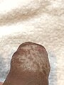
Genital lichen sclerosus of the foreskin

Genital lichen sclerosus of the glans


Possible differential diagnoses of clinical appearance:
- Lichen planus
- Scarring mucosal pemphigoid
- psoriasis inversa
- Vitiligo
- Morphea
- Plasma cell vulvitis or balanitis
- Vulvar or penile intraepithelial neoplasia (VIN / PeIN)
- Squamous cell carcinoma (white skin cancer) of the vulva or penis
Complications
Infections: Bacterial and fungal infections are favored by (mucous) skin defects, which can overlay the disease-specific symptoms
Scarring and adhesions: In women, pseudocysts of the clitoris or vaginal stenosis (narrowing of the vaginal entrance) can result, in men adhesions (sticking) between the glans and the inner sheet of the foreskin, meatal stenosis (narrowing of the urethra orifice) or urethral strictures (narrowing of the urethra). The most common complication in boys and men is phimosis (tightening of the foreskin)
Sensitivity disorders : Even after successful treatment, the patient may continue to experience burning pain or abnormal sensations in the genital area ( vulvodynia or penodynia)
Skin cancer: Compared to the normal population, patients with lichen sclerosus genitalis have an approximately 5% higher risk of developing squamous cell carcinoma of the vulva or penis
diagnosis
During the medical examination, the diagnosis can often be made on the basis of the clinical picture, but in some cases a biopsy (removal of a tissue sample) with a histopathological (light microscopic) examination may be necessary. The British Association of Dermatologists recommends biopsy in the following cases:
- Suspected malignant tumor in long-standing hyperkeratotic, erythematous or abnormally pigmented lesions
- lesions persisting despite therapy
- before initiating any other than standard cortisone therapy (including urological-surgical interventions)
- when performing a circumcision (resection of the foreskin with the resection being sent for histopathological (free examination))
- if extragenital lichen sclerosus is suspected
In children, a biopsy should only be taken in exceptional cases.
An examination for any additional autoimmune diseases is only necessary if there is a corresponding clinical suspicion. Pathogen diagnostics can be carried out in treatment-resistant lesions to clarify possible differential diagnoses or if a secondary infection is suspected.
pathology
Typical histopathological features of lichen sclerosus are atrophy of the epidermis with vacuolar degeneration of the keratinocytes (horn-forming cells of the epidermis) in the basal cell layer and sclerosis of the corresponding dermis. The depth of the sclerosis zone is bordered by an inflammatory infiltrate made up of lymphocytes, which is usually arranged like a ribbon. Other possible changes are irregular acanthosis (widening of the stratum spinosum of the epidermis ) and hyperkeratosis of the epidermis (hypertrophic form), on hairy skin with hyperkeratosis of the hair follicles.
In extragenital lesions, atrophy is often absent and the inflammatory infiltrate can contain eosinophilic granulocytes . In the dermis, dilated small blood vessels are more common. Clinically bullous lesions are associated with dermal edema and even subepidermal blistering.

Lichen sclerosus (atrophic form)

Lichen sclerosus (atrophic form)

Lichen sclerosus (hypertrophic form)

Lichen sclerosus (hypertrophic form)

Lichen sclerosus with follicular hyperkeratosis
.jpg)
.jpg)
.jpg)
.jpg)

Possible differential diagnoses of the histological appearance:
- Lichen planus
- Scarring mucosal pemphigoid
- Mycosis fungoides
treatment
Due to the often chronic course, therapy and aftercare of lichen sclerosus require a long-term concept with the involvement and training of the patient. An early start of therapy should be sought because of the better chances of healing in early lesions.
Medical therapy
A three-month local therapy with highly potent cortisone (e.g. clobetasol propionate 0.05%) as an ointment or cream is recommended for both sexes . Depending on the therapeutic response, this is continued in a reduced dosage and / or application frequency as maintenance therapy in order to reduce side effects typical of cortisone such as thinning of the skin. Local cortisone therapy is also considered safe in the appropriate dosage for children. The success of the therapy should be checked after a period of three months, if necessary with a change of the therapy strategy in the event of non-response and renewed control after a further six months. After that, due to the increased risk of carcinoma, controls should take place at longer intervals if necessary.
In the event of non-response to cortisone as an ointment or cream and in maintenance therapy, the calcineurin antagonists tacrolimus and pimecrolimus can be used locally; steroid injections into single lesions are possible to reduce therapy-resistant itching . Other non-surgical therapy options, which are to be selected depending on the case constellation and side effect profile and which have been tried and tested to varying degrees or are promising, are the local and oral administration of retinoids , antihistamines , methotrexate, fumaric acid , hydroxycarbamide , ciclosporin , TNF-α antagonists and PUVA ( Psoralen plus UV-A).
Surgical therapy
In men with involvement of the foreskin, a circumcision leads to long-term healing in most cases within a maximum of two years postoperatively, possibly after an initial drug therapy attempt. In women, surgical measures for the treatment of lichen sclerosus are not common due to the high risk of recurrence , but functional changes in the urogenital tract may require surgical correction in both sexes. Adhesions, for example between the glans and the inner sheet of the foreskin, can possibly be removed using a laser.
Supportive measures
Fatty ointments and oils support general skin care; the use of skin-irritating perfumes, soaps and shower or personal detergents is not recommended. Excessive mechanical stress on the affected skin areas, e.g. B. by strongly abrasive underwear, possibly also certain sports, can be avoided. In the case of perianal involvement with painful bowel movements, digestive-regulating measures can make bowel movements easier.
Patients should be informed about preventive measures and familial accumulation and, if necessary, instructions on the correct application of ointments or creams should be given, as should awareness of the increased risk of genital squamous cell carcinoma, including the typical signs.
psyche
The itching in the genital area and the resulting compulsion to scratch can be perceived by affected patients as embarrassing. Functional impairments when urinating, defecating or having sexual intercourse reduce the quality of life for both sexes; from a sexual point of view, depending on the severity of the genital skin lesions, cosmetic concerns may arise. Psychosexual complaints can persist even after successful treatment of the disease. Under these circumstances, accompanying psychotherapy or participation in a self-help group can be a useful addition to symptomatic therapy.
literature
- FM Lewis, FM Tatnall, SS Velangi, CB Bunker, A. Kumar: British Association of Dermatologists guidelines for the management of lichen sclerosus, 2018 . In: British Journal of Dermatology . tape 178 , no. 4 , April 2018, ISSN 0007-0963 , p. 839-853 , doi : 10.1111 / bjd.16241 , PMID 29313888 .
- Gudula Kirtschig et al .: European Dermatology Forum Guideline on Lichen sclerosus. (PDF) In: European Dermatology Forum. March 30, 2018, accessed January 5, 2020 .
- Eduardo Calonje, Thomas Brenn, Alexander Lazar, Steven D. Billings: McKee's pathology of the skin with clinical correlations . Fifth ed.Elsevier, no location 2020, ISBN 978-0-7020-7552-0 , p. 486-490 .
Web links
- Gudula Kirtschig: Lichen sclerosus - reason for advice, diagnosis and therapeutic procedure. In: Deutsches Ärzteblatt Online , September 2016
Individual evidence
- ↑ FM Lewis, FM Tatnall, SS Velangi, CB bunker, A. Kumar: British Association of Dermatologists guidelines for the management of union sclerosus, 2018 . In: British Journal of Dermatology . tape 178 , no. 4 , April 2018, ISSN 0007-0963 , p. 839-853 , doi : 10.1111 / bjd.16241 , PMID 29313888 .
- ↑ Arthur Leibovitz, Vladimir Kaplun, Nadya Saposhnicov, Beni Habot: Vulvovaginal examinations in elderly nursing home women residents . In: Archives of Gerontology and Geriatrics . tape 31 , no. 1 , August 2000, p. 1-4 , doi : 10.1016 / S0167-4943 (00) 00059-5 , PMID 10989157 ( elsevier.com [accessed January 4, 2020]).
- ↑ Jenny Powell, Fenella Wojnarowska: Childhood vulvar lichen sclerosus: An increasingly common problem . In: Journal of the American Academy of Dermatology . tape 44 , no. 5 , May 2001, pp. 803-806 , doi : 10.1067 / mjd.2001.113474 , PMID 11312428 ( elsevier.com [accessed January 4, 2020]).
- ↑ William S. Kizer, Troy Prarie, Allen F. Morey: balanitis obliterans Xerotica: Epidemiologic Distribution in on Equal Access Health Care System: . In: Southern Medical Journal . tape 96 , no. 1 , January 2003, ISSN 0038-4348 , p. 9-11 , doi : 10.1097 / 00007611-200301000-00004 , PMID 12602705 ( wkhealth.com [accessed January 4, 2020]).
- ↑ Debra L. Birenbaum, Roger C. Young: High prevalence of thyroid disease in patients with lichen sclerosus . In: The Journal of Reproductive Medicine . tape 52 , no. 1 , January 2007, ISSN 0024-7758 , p. 28-30 , PMID 17286064 .
- ↑ Noritaka Oyama, Ien Chan, Sallie M Neill, Takahiro Hamada, Andrew P South: Autoantibodies to extracellular matrix protein 1 in lichen sclerosus . In: The Lancet . tape 362 , no. 9378 , July 2003, p. 118-123 , doi : 10.1016 / S0140-6736 (03) 13863-9 , PMID 12867112 ( elsevier.com [accessed January 4, 2020]).
- ↑ Adrian Pilatz, Bora Altin Kilic, Eileen Schormann, Lavinia Mägel, Nicole Izykowski: Congenital phimosis in Patients With and Without lichen sclerosus: Distinct expression patterns of Tissue Remodeling Associated Genes . In: Journal of Urology . tape 189 , January 2013, ISSN 0022-5347 , p. 268–274 , doi : 10.1016 / y.juro.2012.09.010 , PMID 23174236 .
- ↑ V. Sherman, T. McPherson, M. Baldo, A. Salim, Xh. Gao: The high rate of familial lichen sclerosus suggests a genetic contribution: an observational cohort study . In: Journal of the European Academy of Dermatology and Venereology . February 2010, doi : 10.1111 / j.1468-3083.2010.03572.x , PMID 20202060 .
- ↑ P. Marren, J. Jell, FM Charnock, M. Bunce, K. Welsh: The association between lichen sclerosus and antigens of the HLA system . In: British Journal of Dermatology . tape 132 , no. 2 , July 29, 2006, p. 197-203 , doi : 10.1111 / j.1365-2133.1995.tb05013.x , PMID 7888355 .
- ↑ Caroline M. Owen, JA Yell: Genital lichen sclerosus associated with incontinence . In: Journal of Obstetrics and Gynecology . tape 22 , no. 2 , January 2002, ISSN 0144-3615 , p. 209-210 , doi : 10.1080 / 01443610120113454 , PMID 12521711 .
- ↑ CB Bunker: Male genital sclerosus and tacrolimus . In: British Journal of Dermatology . tape 157 , no. 5 , November 2007, ISSN 0007-0963 , p. 1079-1080 , doi : 10.1111 / j.1365-2133.2007.08179.x , PMID 17854373 .
- ^ Andreas R. Günthert, Melanie Faber, Gabriele Knappe, Simin Hellriegel, Günter Emons: Early onset vulvar Lichen Sclerosus in premenopausal women and oral contraceptives . In: European Journal of Obstetrics & Gynecology and Reproductive Biology . tape 137 , no. 1 , March 2008, p. 56-60 , PMID 18055095 ( elsevier.com [accessed January 4, 2020]).
- ↑ Eduardo Calonje, Thomas Brenn, Alexander Lazar, Steven D. Billings: McKee's pathology of the skin with clinical correlations . Fifth ed.Elsevier, no location 2020, ISBN 978-0-7020-7552-0 , p. 487 .
- ^ VM Yates, CM King, VK Dave: Lichen sclerosus et atrophicus following radiation therapy . In: Archives of Dermatology . tape 121 , no. August 8 , 1985, ISSN 0003-987X , pp. 1044-1047 , PMID 4026344 .
- ↑ a b c F.M. Lewis, FM Tatnall, SS Velangi, CB Bunker, A. Kumar: British Association of Dermatologists guidelines for the management of lichen sclerosus, 2018 . In: British Journal of Dermatology . tape 178 , no. 4 , April 2018, ISSN 0007-0963 , p. 839-853 , doi : 10.1111 / bjd.16241 , PMID 29313888 .
- ^ HJ Wallace: Lichen sclerosus et atrophicus . In: Transactions of the St. John's Hospital Dermatological Society . tape 57 , no. 1 , 1971, ISSN 0036-2891 , pp. 9-30 , PMID 5570266 .
- ↑ a b Gudula Kirtschig et al .: European Dermatology Forum Guideline on Lichen sclerosus. (PDF) In: European Dermatology Forum. March 30, 2018, accessed January 5, 2020 .
- ↑ Eduardo Calonje, Thomas Brenn, Alexander Lazar, Steven D. Billings: McKee's pathology of the skin with clinical correlations . Fifth ed.Elsevier, no location 2020, ISBN 978-0-7020-7552-0 , p. 489 .
- ↑ JA Carlson, P. Lamb, J. Malfetano, RA Ambros, MC Mihm: Clinicopathologic comparison of vulvar and extragenital lichen sclerosus: histologic variants, evolving lesions, and etiology of 141 cases . In: Modern Pathology: An Official Journal of the United States and Canadian Academy of Pathology, Inc . tape 11 , no. 9 , September 1998, ISSN 0893-3952 , p. 844-854 , PMID 9758364 .
- ↑ A. Larre Borges, D. Tiodorovic-Zivkovic, A. Lallas, E. Moscarella, S. Gurgitano: Clinical, dermoscopic and histopathologic features of genital and extragenital lichen sclerosus: Features of genital and extragenital lichen sclerosus . In: Journal of the European Academy of Dermatology and Venereology . tape 27 , no. November 11 , 2013, p. 1433-1439 , doi : 10.1111 / j.1468-3083.2012.04595.x , PMID 22646723 .
- ↑ Yuri Rojavin, Christopher J. Salgado, Patrick W. Hsu, Jun Liu, James K. Aikins: The surgical management of vulvar lichen sclerosus refractory to medical management . In: Journal of Plastic, Reconstructive & Aesthetic Surgery . tape 61 , no. 7 , July 2008, p. 848-849 , PMID 18439888 ( elsevier.com [accessed January 5, 2020]).
- ↑ Lara J. Burrows, Alia Creasey, Andrew T. Goldstein: The Treatment of Vulvar Lichen Sclerosus and Female Sexual Dysfunction . In: The Journal of Sexual Medicine . tape 8 , no. 1 , January 2011, p. 219–222 , PMID 20955314 ( elsevier.com [accessed January 5, 2020]).
- ^ GL Tasker, F. Wojnarowska: Lichen sclerosus . In: Clinical and Experimental Dermatology . tape 28 , no. 2 , March 2003, ISSN 0307-6938 , p. 128-133 , doi : 10.1046 / j.1365-2230.2003.01211.x , PMID 12653695 .