Neurulation

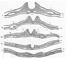
via the neural groove and neural folds
to the neural tube and neural ridges (below)
As Neurulation refers to the formation of the neural tube in chordates and therefore also in humans. The neural tube is the embryonic attachment of the later central nervous system and emerges primarily from the ectoderm by sinking and folding the neuroectoderm .

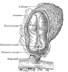
The amniotic sac is open.
(Explanation of the labeling:
Yolk sac = yolk sac , amnion = inner egg membrane , neural groove = neural groove, primitive streak = primitive streak , neurenteric canal = neural groove and yolk sac connecting neurenteric canal , body stalk = sticky stalk ).
Following gastrulation, neurulation begins with the formation of the neural plate , which in the human embryo becomes recognizable on the 19th day of development as a thickening of the ectoderm , as its cells increase in height here. These and other changes are caused by the influence of special signal substances from cells of the axial mesoderm below , the chorda dorsalis , also known as the notochord .
In the case of amphibians, this is preceded by the induction of the Spemann organizer by the Nieuwkoop center , which defines the dorsal (back) side .
Primary neurulation
Phase progression
The process of primary neurulation can be divided into several phases:
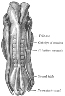
- The beginning of the neurulation can be recognized by the fact that the neural plate is delimited on the surface of the outer germ layer . The ectoderm thickens in the rostral area in front of the primordial mouth (or neurenteric canal ) and the primitive streak.
- The edges of the neural plate bulge out as neural bulges and hold an elongated depression between them, the neural groove . The cells of the midline are attached to the notochord below and thus form the low points of the gutter.
- The neural bulges form into neural folds , which soon unite in the middle and thus close the neural groove, which thus becomes the neural tube . The union of the neural folds is made possible by similar (N-) cadherin molecules in the cell membranes.
- The neuroectoderm then folds away from the rest of the ectoderm, which grows together as a surface ectoderm, and is thus shifted inside the embryo. With this folding of the former Neuralplattenrandes be on both sides of the neural tube, the cells of neural crest formed.
In human embryonic development, the neural tube as an attachment to the central nervous system is folded down from about the 25th day, starting from the middle. Its anterior opening ( neuropore anterior ) closes (with the lamina terminalis ) about a day and a half before the posterior opening ( about 30th day). With this prerequisite, the brain develops from the anterior neural tube section, and the spinal cord that adjoins it at the back . The former cavity of the neural tube is later filled with cerebrospinal fluid as the central canal of the spinal cord or in the inner cerebrospinal fluid spaces of the brain that have expanded to form the ventricular system . One of the first widenings occurs here when the early eye anlage develops as an ocular vesicle.
Induction signals
Neurulation is induced by messenger substances from the notochord, which influence the ectoderm cells above and help determine their interactions. Various protein factors (such as chordin, noggin, follistatin ) suppress the development of the surface epithelium, enable access to developmental genes for nerve tissue and, in conjunction with growth factors, also create regional differentiations.
Molding process
In the middle area of the neural plate, cells of the ectoderm selectively anchor themselves to the notochord - first in a medial line (MHP), then in two dorsolateral formations (DLHP) - and thus become hinge points (HP) for the forming process. Changes in the cell shape can thus be achieved through aligned rearrangements of the cytoskeleton , which in a coordinated manner lead to bulges or retractions in the growing cell network under the lateral pressure of expanding cell layers. Coordinated growth is only possible with the help of the fixed pivot points as an abutment in such a way that - by folding down layers on both sides and connecting in mutual contact - it finally gives shape to the neural tube.
This process is an impressive example of self-organization .
Secondary neurulation
As secondary Neurulation a process is known in which form in a compact cell cord fluid-carrying cavities and flow together into a tubular structure that the lumen connected neural tube and secondarily by neuroepithelium is lined.
In this way, in many vertebrates, a section is attached caudally to the primarily formed neural tube at the level of the 31st somite during the second embryonic month . It is formed in a bulge of mesodermal cells called Eminentia caudalis , the attachment of which goes back to the primitive stripe, and then develops further to the tail area of the spinal cord, which is assigned to the tail . In humans this development is initiated, but it usually does not progress. Relics of the aborted process at the caudal end of the spinal cord are not uncommon (see Filum terminale or Ventriculus terminalis ).
Individual evidence
- ↑ Benninghoff: Macroscopic and microscopic anatomy of humans, Vol. 3. Nervous system, skin and sensory organs . Urban and Schwarzenberg, Munich 1985, ISBN 3-541-00264-6 , p. 79 and p. 105ff.

