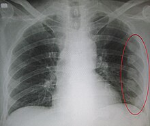
Chest trauma
| Classification according to ICD-10 | |
|---|---|
| S29.9 | Chest trauma |
| S28.0 | Chest crushing |
| S20.2 | Chest contusion |
| ICD-10 online (WHO version 2019) | |
The chest trauma is a violation of the chest , its organs or adjacent structures by mechanical violence from outside or inside. A distinction is made in particular between blunt and penetrating injuries (penetrating the chest wall) and the affected structures (for example bony chest wall , lung tissue, middle layer or blood vessels).
Individual authors also subsume other injuries and damage in the area of the chest such as burns or chemical burns , inhalation trauma due to the inhalation of corrosive, toxic, hot or cryogenic gases and barotrauma under this term. Injuries from chest trauma can be acutely life-threatening.
Epidemiology
| Injuries in multiple trauma patients | |
|---|---|
| Limb injuries | 86% |
| traumatic brain injury | 69% |
| Thoracic trauma | 62% |
| Abdominal injuries | 36% |
| Pelvic injuries | 28% |
| Spinal injuries | 14% |
Chest injuries are a common type of injury. In 45% to 62% of all multiply traumatized patients there are injuries to the chest. After traumatic brain injury , thoracic trauma is the second most common cause of death in multiple trauma patients. In Germany, chest trauma is often an accompanying injury in multiple injuries. A concomitant chest trauma in the accident injured person leads to twice as high a mortality. An additional combination with a traumatic brain injury even quadruples this. The most common injuries are combined skull and extremity injuries, then thorax and extremities, skull and thorax, and then thorax and abdomen . The mortality of multiple trauma patients is mainly determined by traumatic brain and thoracic trauma. Isolated chest injuries are rather rare in German-speaking countries.
history

The trauma is the oldest area of surgery and has its origins in prehistoric people in the first treatment trials of hunting or war injuries. Already here there is evidence of treatment for thoracic injuries. Finds and reports from indigenous peoples show that interventions such as thoracotomies or lung resections were carried out.
In the ancient Egyptian Papyrus Smith , treatments for thoracic injuries are described and these are already classified and divided according to severity and prognosis. There are many reports of thoracic trauma in ancient medical writings , especially war injuries caused by spears or arrows. Even if no therapies for severe thoracic trauma are known here, among others, Hippocrates in the Corpus Hippocraticum gives diagnosis and therapy recommendations for various thoracic diseases, among other things drainage of thoracic fluid accumulations is described for the first time. Different types of dressings are described for chest injuries.
Forms of injury
Typical shapes
As a rule, the term thoracic trauma is used in the literature for injuries caused by mechanical force.
Blunt chest trauma


The blunt chest trauma is the result of a blunt mechanical force from the outside on the thorax. If there are no fractures or injuries to internal organs, it is called a chest bruise .
When the thorax is bluntly violated, the bony structures that make up the thoracic wall are initially affected after the external soft tissues, skin and muscles. The bony chest wall is formed by the thoracic spine , sternum and ribs . In addition to the purely bony injuries, fractures of these structures can lead to further injuries to internal organs through crushing or impaling with sharp-edged bone fragments. Multiple fractures can result in an unstable thorax .
In addition to a lung contusion, injuries to the lungs can also result in a crush injury as lung tears . The latter lead to a pneumothorax , as occurs in 10 to 50 percent of patients with chest trauma, and possibly to a tension pneumothorax . Bleeding into the thorax results in a hemothorax . In a hemopneumothorax , both a pneumothorax and a hemothorax occur in combination. Symptoms are initially shortness of breath and, depending on the extent of the bleeding, circulatory instability . In the case of a pneumothorax, especially when a tension pneumothorax develops, skin emphysema can develop , which can reach a large extent. Therapy is a relief of the pleural cavity by means of chest drainage on the affected side. Smaller intrathoracic bleeding usually stops on its own , so that apart from drainage, no further measures are necessary. A thoracotomy for surgical hemostasis can be performed if there is an initial blood loss of more than 1,500 ml after the chest drainage or if there is continuous blood loss of more than 250 ml per hour for more than four hours.
Injuries to the heart and large vessels, the main artery or the pulmonary arteries , can occur in particular with frontal force on the thorax in combination with rib or sternum fractures. The blunt force of force leads to tissue bruises, tears and hemorrhages. These can damage the tissue in such a way that, in the further course, there may be a complete rupture of vessels or the heart wall or the tearing off of heart structures such as heart valves or papillary muscles . In addition to the risk of bleeding to death , cardiac tamponade (traumatic pericardial tamponade) is a dangerous complication. A rare injury are bronchotracheal injuries such as a tearing or tearing of the windpipe or bronchi . In these cases, an immediate thoracotomy is required to stop the bleeding or to restore and seal the airway.
Penetrating chest trauma

In the case of penetrating chest trauma, the penetration of foreign bodies into the thorax leads to a sharp severing of structures from the chest wall or internal organs. Gunshot and stab wounds are the main cause of penetrating chest injuries.
Hemodynamically stable patients with penetrating chest injuries do not initially require any other acute measures besides chest drainage. In addition to further diagnostics, the focus here is on clinical follow-up, as undetected minor bleeding or injuries to intrathoracic organs can be clinically effective with a delay.
Patients who have a pericardial tamponade after a penetration injury as a result of a heart injury can quickly suffer a circulatory collapse. In addition to a rapid diagnosis using FAST sonography, rapid surgical intervention using an emergency thoracotomy is required. The mortality of patients with penetrating heart injuries is between 35% for stab wounds and 82% for gunshot wounds.
In patients who are circulatory unstable after thoracic penetration injuries, immediate surgical treatment, possibly even as a shock room thoracotomy, is required. Injuries to the heart, aorta or pulmonary vessels are common here. These patients often have a poor prognosis despite immediate emergency thoracotomy. According to various studies, the survival rate is below 20%.
Special forms
These special forms describe injuries and damage in the anatomically defined area of the chest and are not referred to by the majority of authors with the term thoracic trauma .
Barotrauma
In the case of a barotrauma of the lungs, sudden pressure differences between the inside and outside of the thorax can lead to mechanical overloading of the lung tissue or the airways. As a result, the lungs can tear with the formation of a pneumothorax. Smaller tears may not have any consequences. A particular danger is an injury to the lungs as part of a barotrauma involving the pulmonary veins . This can lead to the penetration of small air bubbles into the bloodstream . These can get into the circulatory system via the heart and block smaller arteries there . This can lead to heart attacks or strokes . In addition to decompression sickness accidents while diving , barotraumas of the lungs often occur in patients who are ventilated with positive pressure . With injuries of the airways within the mediastinum can to mediastinalemphysem come.
Inhalation trauma

There are three types of inhalation trauma:
- The thermal inhalation trauma
- The chemical inhalation trauma
- The toxic inhalation trauma
In the case of thermal inhalation trauma, hot gases are inhaled, e.g. B. in fires or explosions or cold gases when inhaling aerosols released cryogenic liquids and substances such. B. liquid nitrogen . Accompanying injuries such as singed hair, burns on the face, soot or burns in the throat or frostbite are external signs. The upper airways and bronchi are mainly affected. Typical symptoms are stridor , cough, shortness of breath and pain.
Chemical inhalation trauma involves inhalation of chemical pyrolysis products, e.g. B. when burning plastics or chemicals. The lower respiratory tract and the lung tissue are damaged. A symptom-free interval of up to 24 hours is possible. This can lead to pulmonary edema and subsequently to acute respiratory distress syndrome . A special form of chemical inhalation trauma is toxic inhalation trauma. This is where toxic gases and combustion products are inhaled. Carbon monoxide poisoning often occurs in fires .
The therapy of inhalation trauma initially consists of stabilizing and securing the vital functions , supplying oxygen and analgesia . In the case of chemical inhalation trauma, in particular, early intubation and ventilation are required , depending on the cause and degree of damage . Toxic substances that have deposited around the mouth and throat can be removed with a simple mouthwash or at least their concentration can be significantly reduced.
Burns
In the case of burns in the thorax area, the general rules for burns initially apply. According to the rule of nine used in burn medicine to determine the surface area of the skin in adults, the thorax takes up 18% of the body surface. There are four degrees of severity:
- Grade 1: There is local redness, swelling and pain in the skin. The epidermis is affected . The injury heals without consequences.
- Grade 2: there is reddening, swelling and blistering at the burn site. The injuries are very painful. The epidermis (grade 2a) or the deeper dermis layers (grade 2b) are affected . Grade 2a injuries heal completely, grade 2b injuries heal with scarring .
- Grade 3: Necrosis occurs , the skin is blackish-white. Because the nerve endings of the skin are destroyed, the injury is often painless. The dermis and subcutaneous tissue are affected . The injuries are irreversible.
- Grade 4: There is deep carbonization of the tissue. Since the nerve endings of the skin are destroyed, the injury is painless. All layers of the skin as well as underlying bones, fascia and organs are affected . The injuries are irreversible.
In addition to the general trauma and vital danger from the burn situation, patients with extensive and circular burns on the thorax are particularly at risk in the acute injury phase. Because tissue that is third to fourth degree burned loses its elasticity, thoracic breathing excursion is restricted. This leads to significantly more difficult or even impossible ventilation . An emergency escharotomy can be life-saving here. Burn injuries to the thorax are often associated with multiple injuries. A combination with an inhalation trauma is possible.
Thoraco-abdominal injuries
Combination injury to the thorax and abdomen is the fourth most common combination in multiple injuries. In particular, the diaphragm, liver and spleen are often also affected in isolated thoracic trauma. In blunt chest trauma, broken ribs in the liver or spleen can spike and cause profuse bleeding. A sudden compression event can cause the diaphragm to rupture on one or both sides and the abdominal organs can be displaced into the chest. This results in a mechanical compression of the lungs with shortness of breath and weakened breathing sounds on the affected side. With a right-sided diaphragmatic rupture, there is a very high probability that there is also a serious injury to the liver. In the case of penetrating chest injuries, foreign bodies such as projectiles or knives that penetrate the body can move into the abdomen and injure organs there. Symptoms of intra-abdominal organ injury are shock and signs of an acute abdomen .
Literature and Sources
- Max Bartels: The medicine of the primitive peoples: Ethnological contributions to the prehistory of medicine. Grieben Verlag, Leipzig 1893. (online)
- Jürgen Durst, Johannes Rohen: Surgical operation theory: in one volume; with topographical anatomy. 2nd Edition. Schattauer Verlag, 1996, ISBN 3-7945-1679-6 , pp. 273-276.
- Ralf Gahr (Ed.): Handbook of Thorax Traumatology. Volumes I and II, Einhorn-Presse Verlag, ISBN 978-3-88756-812-2 .
- Jud W. Gurney et al.: Thorax. Urban & Fischer-Verlag, 2004, ISBN 3-437-23430-7 , pp. 184ff. (on-line)
- Frank Flake et al: 60 cases of emergency services. Urban & Fischer-Verlag, 2006, ISBN 3-437-48230-0 , p. 66ff. (on-line)
- P. Lawin, M. Wendt: Das Thoraxtrauma: Symposium Kassel, February 13 and 14, 1981. Melsungen Bibliomed Med. Verlagsgesellschaft, 1982.
- Guideline "Multiple trauma / treatment of the severely injured". of the DGU at www.awmf.org (accessed on September 19, 2012)
- Axel Rüter among others: trauma surgery. Urban & Fischer-Verlag, 2008, ISBN 978-3-437-21851-4 , p. 35ff. (on-line)
Individual evidence
- ^ K. Ellinger and others: Ellinger, course book emergency medicine. Deutscher Ärzteverlag, 2007, ISBN 978-3-7691-0519-3 , pp. 499-512. (on-line)
- ↑ a b c d e f g h i j k l m n o p q r s t Ralf Gahr (Ed.): Handbuch der Thorax-Traumatologie. Volumes I and II, Einhorn-Presse Verlag, ISBN 978-3-88756-812-2 .
- ↑ a b c d Jürgen Durst, Johannes Rohen: Surgical operation theory: in one volume; with topographical anatomy. 2nd Edition. Schattauer Verlag, 1996, ISBN 3-7945-1679-6 , pp. 273-276.
- ↑ Max Bartels: The medicine of the primitive peoples: Ethnological contributions to the prehistory of medicine. Grieben Verlag, Leipzig 1893. (online) (accessed September 27, 2012)
- ↑ a b c d e guideline "Multiple trauma / treatment of severely injured people". of the DGU at www.awmf.org (accessed on September 19, 2012)
- ^ C. Waydhas, S. Sauerland: Pre-hospital pleural decompression and chest tube placement after blunt trauma: A systematic review. In: Resuscitation. Volume 72, 2007, pp. 11-25.


