Dental plaque
| Classification according to ICD-10 | |
|---|---|
| K03.6 | Deposits (deposits) on the teeth |
| ICD-10 online (WHO version 2019) | |
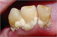
Dental plaque consists of several complex layers and contains proteins , carbohydrates , phosphates and microorganisms . Dental plaque occurs especially where tooth surfaces are not kept free of plaque by natural or artificial cleaning. Plaque can lead to dental caries , periodontal disease, and gingivitis . According to the consistency, a distinction is made between hard (e.g. tartar) and soft plaque (e.g. food residues or bacterial dental plaque such as plaque), and depending on their position on the tooth surface between those located above the gumline (supragingival), and those that are hidden under the gumline and invisible (subgingival).
Emergence
Protein layer
First of all, a precipitate of saliva protein and epithelial flakes forms on the tooth surface (this also includes artificial surfaces such as fillings or dentures ) . This is called Pellicle in the English technical literature . Pellicle forms a thin protective layer within about half an hour and can be rinsed off. In contrast, the plaque and the cuticle, the skin of the teeth, can only be removed with toothbrushes. Removal of the cuticle is not necessary for dental health.
Bacterial colonization
On this protein layer, which is only a few micrometers thick, bacteria that belong to the normal oral flora can settle with the help of the mucous parts of the saliva ( mucins ) ( according to current knowledge, Streptococcus mutans is not part of the normal bacterial flora of the oral cavity). Streptococcus mutans forms dextrans , which contribute to the formation of plaques. These microorganisms have special receptors on their cell wall that enable them to bond. This prevents them from being flushed into the stomach , which would mean certain death.
Symbiosis of bacteria
If this process can run undisturbed, new microorganisms settle on the first layer of bacteria and multiply. According to the findings of biofilm research, the bacteria do not simply stick to one another, but rather form a symbiosis in which they supply one another with metabolic products . Special contact molecules stabilize the bacterial community. Channels that allow the diffusion of substances run within the bacterial layer . A matrix of protein and carbohydrates forms between the bacteria , which serves as a food reserve and mechanically strengthens the layer.
Biofilms have a tough life, and the use of antiseptic mouthwashes can only damage the upper cell layer. Since bacteria only need half an hour to divide, this layer is restored within a short time.
consequences
These microorganisms are favored with high and frequent sugar consumption . This leads u. a. to acid formation , with it to carious lesions and finally to tooth decay . The dental plaque, d. H. the matrix, consisting of layers of leftover food, living microorganisms and their metabolic products, absorbs minerals from the saliva after a few hours and can harden into tartar . Tartar is rougher than the natural tooth surface (or polished fillings) and encourages new bacterial colonization. Its removal is therefore indicated by tartar removal or as part of a professional tooth cleaning (PZR).
Certain ( anaerobic ) microorganisms also produce substances that irritate the immune system . This leads to inflammation of the gums ( gingivitis ). The irritation causes swelling and reddening of the gums, which bleed easily when touched. If the inflammation continues in sensitive people, periodontitis can develop. Then tartar can also develop below the gum line, which contains minerals from blood and gum secretion (has a different composition than the tartar above the gum line, which is mineralized by saliva components).
In addition to periodontitis and tooth decay, bacteria also form odorous sulfur compounds in plaque , which is what causes bad breath .
Determination of bacterial plaque (plaque test)
To make the plaque on the tooth surfaces and the oral mucosa visible, coloring tablets or solutions are used. These are also known as "plaque indicators" or "plaque levelers".
The test discolors the plaque and shows where the teeth have not yet been adequately cleaned. Various plaque stains are used here.
Solid color
Tablets with erythrosine stain areas of plaque on the teeth and the oral mucosa . The dye, which has a high iodine content but is approved as a food coloring, is suspected of causing allergies and should therefore not be used over the long term. See also iodine intolerance .
Two-tone staining
The test differentiates between older and newer plaque by means of various color additives. More neglected areas on the tooth become visible and can be cleaned more thoroughly in the future. The coloring tablets contain brilliant blue ( CI 42090) and phloxin B (CI 45410) as coloring agents . Phloxin (tetrachlorotetrabromofluorescein) is one of the xanthene dyes.
UV light
This rinsing solution specially developed for the dental practice contains fluorescein . Dental plaque fluoresces under UV light . This coloration remains invisible in normal light. When used properly, no health risks are to be expected.
Previously used solutions with the dyes fuchsine or crystal violet may contain amines that are harmful to health due to the manufacturing process . Continuous use of large amounts poses a carcinogenic risk.
Elimination (teeth cleaning)
Fresh plaque can be removed mechanically by brushing your teeth. As soon as the soft plaque turns into plaque due to mineralization , plaque can no longer be removed simply by thorough mechanical cleaning with a toothbrush and toothpaste . An effective mechanical cleaning process uses ultrasound processes or removal with dental hand instruments ( scaler ).
prophylaxis
Sustainable cleaning can only be achieved through thorough daily cleaning. Some chemical agents (e.g. chlorhexidine solutions) can inhibit the formation of new plaque (but not remove plaque). The combination of home oral hygiene, professional oral hygiene and consistent adherence to control intervals can prevent both the onset and recurrence of oral cavity diseases.
literature
- Philip Marsh, Michael V. Martin: Oral Microbiology. Thieme, Stuttgart 2003, ISBN 3-13-129731-X . (Translation into German of the 4th edition in English Oral Microbiology. Reed Educational and Professional Publishing Ltd., 1999)
- CJ Adler, K. Dobney, et al. a .: Sequencing ancient calcified dental plaque shows changes in oral microbiota with dietary shifts of the Neolithic and Industrial revolutions. In: Nature Genetics . Volume 45, Number 4, April 2013, ISSN 1546-1718 , pp. 450-455, 455e1. doi : 10.1038 / ng.2536 . PMID 23416520 .
Individual evidence
- ↑ a b c D. Heidemann: Periodontology . Urban and Schwarzenberg, 1997, ISBN 3-437-05490-2 .
- ↑ Ph. Marsh, MV Martin: Orale Mikrobiologie. 1st edition. Thieme Verlag, 2003, ISBN 3-13-129731-X , p. 86.
- ↑ Ph. Marsh, MV Martin Orale Microbiology. 1st edition. Thieme Verlag, 2003, ISBN 3-13-129731-X , p. 49.
- ↑ S. Schellerer: Intensive care for radiant teeth . In: Pharmaceutical newspaper. 42/2008.
- ^ S. Paraskevas, MM Danser, MF Timmerman, U. Van der Velden, GA van der Weijden: Optimal rinsing time for intra-oral distribution (spread) of mouthwashes. In: Journal of Clinical Periodontology . Volume 32, Number 6, June 2005, ISSN 0303-6979 , pp. 665-669. doi : 10.1111 / j.1600-051X.2005.00731.x . PMID 15882228 .
- ↑ P. Axelsson, B. Nyström B., J. Lindhe .: Long-term effect in biofilm management on tooth preservation, caries formation and periodontal diseases in adults. Results after 30 years of investigation . In: Journal of Clinical Periodontology . 2004 Sep; 31: 749-57.

