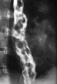
Esophageal varices
| Classification according to ICD-10 | |
|---|---|
| I85 | Esophageal varices |
| ICD-10 online (WHO version 2019) | |
Esophageal varices are varicose veins ( varices ) of the esophagus ( esophagus ). These expansions of the venous plexus in the wall of the esophagus are the result of a disruption of the venous blood circulation and mostly caused by portal hypertension , for example as a result of liver cirrhosis . Bleeding from esophageal varicose veins is a life-threatening complication and a medical emergency.
causes
The nutrient-rich but oxygen-poor venous blood from the spleen, stomach, intestines and gallbladder normally flows via the portal vein through the liver and from there into the inferior vena cava. If this blood flow is restricted (e.g. due to cirrhosis of the liver ), portal hypertension occurs (increased blood pressure in the portal vein). In this case, the liver is bypassed via the portocaval anastomoses (connections between the portal vein and the upper and lower vena cava), and the blood then flows directly into the vena cava.
There are several such portocaval anastomoses: in the rectum , in the abdominal wall, in the stomach, and in the esophagus . The latter widen to esophageal varices when the blood pressure in the portal vein is increased. Bleeding from these varices can be life threatening.
Epidemiology (frequency)
In severe portal hypertension, for example in the context of liver cirrhosis , around half of those affected have esophageal varices. The lethality of a bleeding is up to 30% even with treatment. The probability of a relapse following the first esophageal variceal bleeding is around 70%.
Complications
The dilated veins in the lower end of the esophagus , the esophageal varices, can, because they are very thin-walled, tear easily and lead to profuse bleeding. The blood loss, if severe enough, leads to shock and can be life-threatening. This bleeding is often complicated by an existing bleeding disorder caused by cirrhosis of the liver . Slight bleeding leads to tarry stools (melena), acute life-threatening bleeding typically leads to vomiting of blood ( hematemesis ). In addition, other complications such as ascites and hepatic coma can occur in patients with cirrhosis, independent of bleeding . One study showed that treatment for hepatic encephalopathy can also lower the risk of other cirrhosis complications such as spontaneous bacterial peritonitis (SBP) or variceal bleeding .
diagnosis
The diagnosis is made endoscopically by gastroscopy . If there is bleeding, an attempt to stop the bleeding can also be undertaken as part of a gastroscopy. The main purpose of gastroscopy is to answer the question of whether there are other sources of bleeding. Esophageal varices are clinically important primarily as a source of bleeding. Other sources of bleeding in the stomach or esophagus are Mallory-Weiss syndrome , ulcer bleeding and gastric mucosal erosions .
Staging
According to the appearance and characteristics during the endoscopy , clinical stages can be classified into grades I - IV:
- Stage I: The submucosal veins are widened, but they disappear after air insufflation through the endoscope.
- Stage II: There are individual varices protruding into the lumen of the esophagus, which persist even with air insufflation.
- Stage III: The lumen of the esophagus is narrowed by protruding variceal cords. As a sign of epithelial damage ( erosion ), reddish spots ( "cherry spots" ) can exist on the mucous membrane.
- Stage IV: The variceal cords have obstructed the esophageal lumen and there are usually numerous erosions of the mucous membrane.
In addition to esophageal varices, some of the patients also have gastric varices and gastropathia hypertensiva .
therapy
In therapy, a distinction must be made between acute measures in the event of bleeding and prophylaxis against bleeding or prophylaxis against recurrence.
Therapy for acute bleeding
In an emergency, the affected patient should be placed directly in an intensive care unit. The primary goal is hemostasis. This can best be achieved by means of a rubber band ligation of the bleeding varices, injection of Histoacryl ( N-butyl cyanoacrylate ) or variceal obliteration by injection.
If endoscopic treatment of the varices is not possible, a balloon tube should be used to stop bleeding by means of compression, e.g. B. the Sengstaken-Blakemore probe or the Linton-Nachlas probe . The patient should then be transferred to endoscopic therapy as soon as possible. Until sclerosis or tamponade through probes, the portal venous blood flow can be reduced by the administration of terlipressin or (off-label) somatostatin or octreotide .
Other general measures are:
- Monitoring of vital functions
- Endotracheal intubation ( risk of aspiration )
- Volume administration via large-lumen peripheral venous access
- Administration of antibiotics (threatened sepsis )
- Management of the underlying disease (e.g. cirrhosis of the liver )
Therapy to prevent relapse or bleeding
The underlying cause of portal hypertension is to be treated as causal therapy. However, not all causes of portal hypertension can be treated, so that often only symptomatic and delaying therapy is used.
Drug therapy includes the administration of beta blockers , nitrates and spironolactone to lower the pressure in the portal circulation . Ligation treatment is the method of choice because serious complications rarely occur.
Interventional and surgical procedures usually aim to create a shunt between the portal vein circulation and the systemic venous circulation. Since the liver is bypassed and thus loses its ability to metabolize toxins such as ammonia , shunts often increase the likelihood of further complications of liver cirrhosis , such as portal hypertension or hepatic encephalopathy . Common procedures are:
- TIPS (Transjugular Intrahepatic Portosystemic Shunt)
- Shunt operations:
- Portocaval Shunt (Portocaval End-to-Side Anastomosis, PCA)
- Splenorenal shunt


