Chlamydophila pneumoniae
| Chlamydophila pneumoniae | ||||||||||
|---|---|---|---|---|---|---|---|---|---|---|

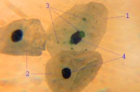
Chlamydophila pneumoniae in epithelial cells : 1 - infected epithelial cell, 2 - uninfected epithelial cells, 3 - C. pneumoniae as reticular bodies in the cell, 4 - cell nuclei |
||||||||||
| Systematics | ||||||||||
|
||||||||||
| Scientific name | ||||||||||
| Chlamydophila pneumoniae | ||||||||||
| ( Grayston et al., 1989) Everett et al., 1999 |
Chlamydophila pneumoniae (formerly known as Chlamydia pneumoniae ) are bacteria that infect humans and can cause pneumonia, among other things. They are a type of chlamydia . Chlamydophila pneumoniae has a complex life cycle and must infect another cell in order to reproduce itself. In addition to its role in pneumonia, there is evidence that the pathogen can be associated with arteriosclerosis and asthma . The full DNA sequence of the genome was published in 1999.
Life cycle and methods of infection
Chlamydophila pneumoniae is a small gram-negative bacterium that goes through several transformations in its life. It exists as an elementary body (EB) between biological host systems. The EB is not metabolically active, only 0.2-0.3 micrometers in size, but is resistant to the environment (e.g. osmotic changes) and can also survive outside the host system. The EB travels from an infected person to the lungs of an uninfected person in small droplets and causes the infection. Once in the lungs, the EB is absorbed by ( epithelial ) cells in a kind of bag (membrane enclosure) called an endosome , a process known as endocytosis . However, the EB is not destroyed by fusion with the lysosomes , which is typical of phagocytosed material. Instead, it turns into a reticular body (RB), one micrometer in size, and begins to multiply within the endosome. The RBs must use some of the host cell's machinery to complete their reproduction. The reticular bodies then develop back into elementary bodies and are released back into the lungs, often causing the host cell to die. The EBs are then able to infect new cells, either in the same organism or in a new one. This is why the life cycle of Chlamydophila pneumoniae is divided into EB, which can only cause infections but cannot multiply, and RB, which can multiply but cannot cause new infections.
In addition to these two main forms of the development cycle, another form is known, the aberrant corpuscles. This is an intracellular, persistent form that arises under non-optimal growth conditions of the host cells. It is viewed as a permanent form with a reduced metabolism and can develop back into elementary bodies. The aberrant bodies are also of medical importance as their presence has been linked to reactive arthritis.
Pneumonia , arthritis, tendonitis due to Ch. Pneumoniae
Chlamydophila pneumoniae is a common cause of pneumonia in people with compromised immune systems worldwide. Chlamydophila pneumoniae is typically transmitted by air (through the air). Most infections proceed without any discomfort or in the form of a slight sore throat. The chlamydia can lead to post-infectious arthritis and tendonitis four to six weeks after the primary infection. Usually neither the doctor nor the patient establishes a connection between the possibly symptomatic primary infection and the joint pain. It is assumed that the population is 50-70% infected with Chlamydophila pneumoniae . This high level of contamination indicates a lingering in the organism for several years. Because treatment and diagnosis have historically been different, terms such as Chlamydia pneumoniae are used and one speaks of atypical pneumonia.
Symptoms and diagnosis
Most infections are asymptomatic and unnoticed or cause a slight sore throat, but they can also be the cause of bronchitis (also an exacerbation of bronchial asthma) or otitis media and, rarely, Guillan-Barré syndrome and endocarditis . If pneumonia occurs in a weakened person, symptoms of an infection with Chlamydophila pneumoniae cannot be differentiated from those of other pneumonia (cough, fever and shortness of breath). Chlamydophila pneumoniae are more likely to cause sore throats, hoarseness (from laryngitis or pharyngitis), and sinusitis than other types of pneumonia, but other types of pneumonia can also have these symptoms. A distinction is therefore not possible. Usually, a doctor is also unable to make a clear diagnosis based on symptoms alone.
The diagnosis of Chlamydophila pneumoniae can be made by examining the sputum or secretions of the throat. Such an examination is not used in practice because of the high probability of a false negative result, since the number of detectable chlamydia is very low. A blood test can show antibodies to the bacterium. However, if there was a previous infection, the antibodies could also come from it. Therefore an analysis of the antibodies is necessary after 6 weeks in order to diagnose a new or old infection. The blood test can also show proteins (antigens) of Chlamydophila pneumoniae , either by direct fluorescent antibody testing , also called enzyme-linked immunosorbent assay ( ELISA ), or by polymerase chain reaction ( PCR ).
X-rays of the lungs infected with Chlamydophila pneumoniae often show a small patch of heightened shadow (opacity). However, there are also different patterns here, so that a clear analysis is often not possible.
Treatment and Chances of Success
Typically, treatment begins before the microorganism has been identified, at least if a known clinical picture is recognizable. This general therapy includes an antibiotic against atypical bacteria, which includes Chlamydophila pneumoniae . Since many pneumonia and inflammation of the arterial system z. B. run without severe symptoms, it is usually only possible to find a suitable therapy after more detailed examinations of the secretion. Because of the problem of the different states of Chlamydophila pneumoniae , a different form of therapy is necessary for each phase, as it is pointless to use an antibiotic against the reticular body that does not work against the elementary form, otherwise the elementary bodies will revert to their own after weaning RB change and the disease starts all over again. Therefore, in the future there will have to be an effective antibiotic therapy in the form of a combination agent, at least a combination that also includes the dormant forms.
The most commonly used antibiotics are macrolides such as azithromycin or clarithromycin . If a test clearly shows Chlamydophila pneumoniae as the cause, therapy with doxycycline can be continued, which has fewer side effects and is somewhat more effective against this bacterium. Sometimes a quinolone antibiotic such as levofloxacin or moxifloxacin is also used to start therapy, but this has more side effects. Treatment usually lasts 10 to 14 days. In post-infectious arthritis or tendinitis, Chlamydophila pneumoniae is most efficiently inhibited in vitro by clarithromycin. Here antibiotic therapy must be carried out for 30–90 days.
β-lactam antibiotics such as penicillins are ineffective due to the lack of a cell wall in these bacteria. On the contrary, penicillin could interrupt the development cycle of chlamydia and make them persistent.
The prognosis for pneumonia from Chlamydophila pneumoniae is good. Hospital stays are atypical, complications are rare and most patients have no after-effects after recovering from illness. However, the reticular corpuscles or aberrant corpuscles usually survive in the infected macrophages, or at least the initial symptoms disappear for the time being. However, if Chlamydophila pneumoniae becomes chronic, i.e. the body does not manage to eliminate the infection itself, the risk of creeping diseases of the circulatory system, nervous system, etc. is considerable.
Epidemiology and preparedness
Chlamydophila pneumoniae affects all age groups and is most common in the group of 60 to 79 year olds. Re-infection after a short period of immunity is common. In younger people, the infection often persists (high degree of contamination), but usually does not appear for a long time. After a long time z. B. but a chronification of Chlamydophila pneumoniae result, which then manifests itself again in the different forms ( arthritis , arteriosclerosis, etc.). The contamination is estimated at 50–70%. The incidence of pneumonia is one in a thousand people a year and affects around ten percent of all pneumonia without hospitalization.
For diseases of the circulatory system, however, according to the research results of the last few years, it can be assumed that a considerably larger number can be attributed to Chlamydophila pneumoniae . Arteriosclerosis in particular and its consequences (heart disease, strokes, etc.) is possibly primarily induced by Chlamydophila pneumoniae , as more and more studies indicate.
In clinical cases, Chlamydophila pneumoniae is becoming more and more resistant to therapy , including doxycycline and erythromycin .
There is currently (2016) no effective vaccination. Apart from stringent hygiene measures and avoiding exposure, there are no preventive measures.
asthma
Bronchial asthma and exacerbations of this disease are often associated with the serological detection of Chlamydia pn. The symptoms can respond well to therapy with macrolides .
Other diseases that Chlamydophila pneumoniae causes
In addition to pneumonia, Chlamydophila pneumoniae causes other diseases. These include meningoencephalitis , arthritis , BOOP ( bronchiolitis obliterans with organizing pneumonia), myocarditis or endocarditis and Guillain-Barré syndrome , which often require a longer duration of therapy. It has also been linked to dozens of other diseases, such as Alzheimer's disease , multiple sclerosis , fibromyalgia , chronic fatigue syndrome , prostate problems, and many others.
Links between Ch. Pneumoniae and chronic diseases
In addition to known acute infections, Chlamydophila pneumoniae has been linked to other chronic diseases. There is some evidence that the onset of asthma may be related to Chlamydophila pneumoniae . However, until 2005 there was no proof.
Links to Chlamydophila pneumoniae infections and heart attacks have been found. In fact, the bacterium has been detected in coronary arteries, among others. Antibodies are also higher in people with heart problems. In stroke patients, too, signs of a chronic infection with Chlamydophila pneumoniae were repeatedly found. It is assumed that chronic infections with the pathogen promote the development and progression of arteriosclerosis and thus play a role in heart attacks and strokes . Indian researchers and physicians at the University of Munich have meanwhile found that heart attacks associated with Chlamydophila pneumoniae decreased after the administration of suitable antibiotics and that even narrowing of the coronary arteries regressed.
literature
- S. Kalman et al. (1999): Comparative genomes of Chlamydia pneumoniae and C. trachomatis. Nature Genetics 21: pp. 385-389, PMID 10192388
- S. O'Connor et al .: Potential Infectious Etiologies of Atherosclerosis: A Multifactorial Perspective . Emerging Infectious Diseases , Vol. 7, Sept. – Oct. 2001
- DL Hahn, RW Dodge, R. Golubjatnikov: Association of Chlamydia pneumoniae (TWAR) infection with wheezing, asthmatic bronchitis and adult-onset asthma. JAMA 1991, 266: pp. 225-230.
- CP Cannon, E. Braunwald, CH McCabe, JT Grayston, B. Muhlestein, RP Giugliano, R. Cairns, AM Skene: Pravastatin or Atorvastatin Evaluation and Infection Therapy-Thrombolysis in Myocardial Infarction 22 Investigators. Antibiotic treatment of Chlamydia pneumoniae after acute coronary syndrome. N Engl J Med . 2005 Apr 21; 352 (16): pp. 1646-1654, PMID 15843667
- Klaus-Armin Bartsch: Representation of activated NF-κB in the arteriosclerotic lesion.
- Marianne Abele-Horn: Antimicrobial Therapy. Decision support for the treatment and prophylaxis of infectious diseases. With the collaboration of Werner Heinz, Hartwig Klinker, Johann Schurz and August Stich, 2nd, revised and expanded edition. Peter Wiehl, Marburg 2009, ISBN 978-3-927219-14-4 , p. 190.
Web links
Individual evidence
- ↑ Helmut Hahn, Stefan HE Kaufmann, Thomas F. Schulz, Sebastian Suerbaum (eds.): Medical microbiology and infectious diseases. 6th edition, Springer Verlag, Heidelberg 2009, ISBN 978-3-540-46359-7 , pp. 416-426.
- ↑ Marianne Abele-Horn (2009), p. 190.
- ^ KM Sandoz, DD Rockey: Antibiotic resistance in Chlamydiae. In: Future microbiology. Volume 5, number 9, September 2010, pp. 1427-1442, doi : 10.2217 / fmb.10.96 , PMID 20860486 , PMC 3075073 (free full text) (review).
- ^ ES Gold, RM Simmons, TW Petersen, LA Campbell, CC Kuo, A. Aderem: Amphiphysin IIm is required for survival of Chlamydia pneumoniae in macrophages. In: The Journal of experimental medicine. Volume 200, number 5, September 2004, pp. 581-586, doi : 10.1084 / jem.20040546 , PMID 15337791 , PMC 2212749 (free full text).
- ↑ L. Richeldi, G. Ferrara, LM Fabbri et al .: Macrolides for chronic asthma (Cochrane Review). The Cochrane Library, Issue 1. Oxford 2003