Nociceptor
A nociceptor (of lat. Nocere , damage ') - also known as Nozisensor or incorrectly as Nozirezeptor denotes - is a free sensory nerve ending, the event of an imminent or carried tissue damage (by thermal, chemical or mechanical noxae ) electrical signals ( action potentials ) generated . Nociceptors form the starting point of nociception , their irritation is thus typically accompanied by a sensation of pain .
Medical history
The term nociceptor was coined in 1906 by the British physiologist Charles Scott Sherrington . In the 1960s, the American physiologist Edward Perl (see web links) made significant contributions to the elucidation of the function of nociceptors.
description
Depending on the type of nerve fiber: Aδ fiber (sparsely myelinated, diameter 1–6 µm, conduction velocity 5–30 m / s) or C fiber (non-myelinated, diameter 0.2–1.5 µm, conduction velocity 0.5–2 m / s) and their specific reactivity, nociceptors can be divided into three groups:
- Mechanonociceptors that respond to strong, especially sharp stimuli (endings of Aδ fibers)
- Polymodal nociceptors that also react to heat and cold pain and chemical pain stimuli. Depending on the fiber properties, they can be differentiated into Aδ-polymodal nociceptors and C-polymodal nociceptors .
- Silent nociceptors , nociceptors that cannot be excited in healthy tissue, the stimulus threshold of which is lowered to a particularly sensitive level by inflammation (endings of C fibers).
The first type is responsible for triggering the first pain and protective reflexes, whereas the second type characterizes the longer lasting, delayed pain (usually burning over an area).
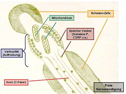
In the skin there are nociceptors as free nerve endings , so-called intraepidermal nerve fiber endings [IENFE], of afferent Aδ and C fibers in a ratio of 1: 7. Free nerve endings are characterized by a peripheral branch. They have a fenestrated covering made of Schwann cells and have numerous varicosities (swellings). These are mostly located near blood vessels and mast cells . Some of them penetrate the epidermis . Nociceptors are of crucial importance for the properties of the skin as a protective covering for the organism. In certain diseases (e.g. diabetes mellitus ), the nociceptors at the ends of the longest nerve fibers (in the feet) can wither; the result is insensitivity to pain in injuries to the feet.
The density (i.e. number per area) of nociceptors in humans is greater than that of any other skin receptor . The distribution of the free IENFE on the body surface is not uniform and varies considerably between individuals (by a factor of 10) between healthy normal persons. The density of the heat nociceptors - determined in 8 body regions - is highest at the fingertips, followed by the palms, forehead, soles of the feet, shoulders, back, calves, and lowest at the dorsum of the feet. You can find nociceptors in the muscles, around the intestines and other parts of the body. Ligaments and tendons of the joints also contain nociceptors, e.g. B. on the foot, however, per area about 100 times less than in the skin.
In the human body there are nociceptors in almost every tissue. Exceptions include visceral organs such as the brain , parenchyma of the liver , lungs , the inner leaf of the lung membrane (= pleura pulmonalis, = pleura visceralis), kidney and adrenal glands , spleen , but also thyroid , pancreas , cartilage , inner parts of the intervertebral discs , synovial membrane , Ligamentum anterius and vertebral body (with the exception of the posterior quarter), uterine body and visceral peritoneum (peritoneum) , visceral pleura (lung membrane ), parts of the pericardium (visceral pericardium) as well as retina , vitreous humor, eye lens and tooth enamel , which themselves have no or no significant accumulation have nociceptors, but are usually encased by one or more pain-sensitive tissues or connective tissue capsules (e.g. the meninges and the liver capsule or the parietal peritoneum and the parietal pleura ), from which (sometimes enormous) pain can arise.
For mammals, it has been proven that nociceptors are embodied by so-called free nerve endings of the thin, sensitive Aδ and C nerve fibers. The excitation by thermal, mechanical or chemical pain stimuli occurs at high stimulus intensities , see pain threshold , see quantitative sensory testing . Characteristic is little or no adaptive control .
Substances that activate nociceptors are called algogenic , these substances cause pain. Algogenic substances are z. B. serotonin , bradykinin , histamine as well as potassium ions and leukotrienes . Formaldehyde is also used in pain research .
ontogenesis
In embryonic development, nociceptors, like nerve tissue, generally arise from the ectoderm .
See also
literature
- Introduction: Nociception and Pain (PDF; 1.4 MB)
Web links
- Edward Perl , studied how nociceptors work.
Individual evidence
- ^ Flexicon: Nociception
- ↑ Nociception ( Memento of the original from March 21, 2016 in the Internet Archive ) Info: The archive link was inserted automatically and has not yet been checked. Please check the original and archive link according to the instructions and then remove this notice.
- ↑ Lauria G, Holland N, Hauer P, Cornblath DR, Griffin JW, McArthur JC .: Epidermal Innervation: Changes With Aging, Topographic Location, and in Sensory Neuropathy. In: J Neurol Sci. 1999, pp. Vol. 164, 172-178 , accessed June 18, 2020 .
- ↑ V. Provitera, CH.Gibbons, G.Wendelschafer-Crabb, V. Donadio, DF.Vitale, A.Stancanelli, G.Caporaso, R.Liguori, N.Wang, L.Santoro, WR.Kennedy, M.Nolano : A multi-center, multinational age- and gender-adjusted normative dataset for immunofluorescent intraepidermal nerve fiber density at the distal leg . In: European Journal of Neurology . tape 23 , 2016, p. 333-338 , doi : 10.1111 / ene.12842 .
- ↑ F.Mancini, A. Bauleo, J. Cole, F.Lui, CA. Porro, P. Haggard, GD Iannetti: Whole-body mapping of spatial acuity for pain and touch . In: Annals of Neurology . tape 75 , 2014, p. 917-924 , doi : 10.1002 / ana.24179 .
- ↑ S. Rein, U. Hanisch, H.Zwipp, A.Fieguth, S.Lwowski, E. Hübers: Comparative analysis of inter- and intraligamentous distribution of sensory nerve endings in ankle ligaments. A cadaver study . In: Foot Ankle Int . tape 34 , 2013, p. 1017-1024 , doi : 10.1177 / 1071100713480862 .
- ↑ S.Rein, S.Manthey, H.Zwipp, A.Witt: Distribution of sensory nerve endings around the human sinus tarsi: a cadaver study . In: Journal of Anatomy . tape 224 , 2014, p. 499-508 , doi : 10.1111 / joa.12157 .
- ^ Christian Rieger, Horst von der Hardt, Felix Hans Sennhauser, Ulrich Wahn, Maximilian S. Zach: Pediatric Pneumology . Springer-Verlag, 2013, ISBN 978-3-662-09182-1 , pp. 25 ( limited preview in Google Book Search [accessed February 4, 2017]).
- ↑ HA Baar, HU Gerbershagen: pain, pain disease, pain clinic . Springer-Verlag, 2013, ISBN 978-3-642-71220-3 , pp. 18 ( limited preview in Google Book Search [accessed February 4, 2017]).
- ↑ Lois White, Gena Duncan, Wendy Baumle: Medical Surgical Nursing: An Integrated Approach . Cengage Learning, 2012, ISBN 1-4354-8802-4 , pp. 874 ( limited preview in Google Book Search [accessed June 28, 2016]).
- ^ Brian Duvall, Al Lens, Elliot B. Werner: Cataract and Glaucoma for Eyecare Paraprofessionals . SLACK Incorporated, 1999, ISBN 978-1-55642-335-2 , pp. 4 ( limited preview in Google Book Search [accessed June 28, 2016]).
- ↑ Susanne Geyer, Arthur Grabner: Die Tierarzthelferin: Textbook and guide for training to become a veterinary assistant . Schlütersche, 2005, ISBN 978-3-87706-586-0 , p. 195 ( limited preview in Google Book Search [accessed May 7, 2016]).
- ↑ Florian Lang, Philipp Lang: Basic knowledge of physiology . Springer-Verlag, 2007, ISBN 978-3-540-71402-6 , pp. 380 ( limited preview in Google Book Search [accessed May 7, 2016]).
- ↑ Bernd Hartmann, Michael Spallek, Rolf Ellegast: Work-related musculoskeletal disorders: causes, prevention, ergonomics, rehabilitation . ecomed-Storck GmbH, 2013, ISBN 978-3-609-16478-6 , p. 49 ( limited preview in Google Book Search [accessed May 7, 2016]).
- ↑ Grünwald Frank, Derwahl Karl-Michael: Diagnosis and Therapy of Thyroid Diseases: A Guide for Clinic and Practice . Lehmanns Media, 2014, ISBN 978-3-86541-538-7 , pp. 93 ( limited preview in Google Book Search [accessed May 23, 2016]).
- ^ Peter Altmeyer, Torsten Liem, Angela Schleupen, René Zweedijk: Osteopathic treatment of children . Georg Thieme Verlag, 2012, ISBN 978-3-8304-7605-4 , p. 861 ( limited preview in Google Book Search [accessed June 28, 2016]).
- ↑ Roland Schiffter, Elke Harms: Connective tissue massage: neural processes - findings - practice . Georg Thieme Verlag, 2009, ISBN 978-3-13-152455-3 , p. 67 ( limited preview in Google Book Search [accessed June 28, 2016]).
- ↑ Winfried Mohr: Joint pathology: Historical bases, causes and developments of joint diseases and their pathomorphology . Springer-Verlag, 2013, ISBN 978-3-642-57071-1 , pp. 125 ( limited preview in Google Book Search [accessed June 28, 2016]).
- ↑ D. Rosenow, V. Tronnier, H. Göbel: Neurogenic pain: Management of diagnostics and therapy . Springer-Verlag, 2005, ISBN 978-3-540-26483-5 , pp. 7 ( limited preview in Google Book Search [accessed February 4, 2017]).
- ^ Glossary , University of Bern
- ↑ Peter Reuter: Springer compact dictionary medicine. Concise Medical Dictionary: German-English. Springer, 2005. ISBN 3-540-23780-1 , p. 724.
- ↑ Wolf Erhardt: Anesthesia and analgesia in small and domestic animals as well as in birds, reptiles, amphibians and fish. Schattauer Verlag, 2004, ISBN 3-7945-2057-2 , p. 405.
