Gamma Spectroscopy

Gamma spectroscopy is the measurement of the spectrum of the gamma radiation from a radioactive radiation source . Gamma quanta do not have arbitrary, but rather certain ( discrete ) energies that are characteristic of the respective radionuclide , similar to the way in which the spectral lines are characteristic of the substances contained in the sample in optical spectroscopy . That is why gamma spectroscopy is an important method for examining radioactive substances, for example radioactive waste, in order to be able to decide on their treatment.
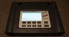
Some gamma spectrometers are commercially available under the name Radionuclide Identifying Device . These are devices for identifying a gamma emitter, not for quantitative measurement of activity.
One can more precisely distinguish between the
- Gamma spectroscopy , which shows as a qualitative measurement which nuclides are present,
- and gamma spectrometry , which quantifies the activity of the individual nuclides.
However, the terms are not used in a completely uniform way. The device is generally called a gamma spectrometer, not a “gamma spectroscope”.
Structure of a gamma spectrometer
detector

The main part of the measuring apparatus, the gamma spectrometer , is a suitable radiation detector . For most gamma emitters with energies between about 50 keV and a few MeV , semiconductor detectors made of high purity germanium ( high purity germanium , abbreviation HPGe) or less pure, lithium doped ("drifted") germanium (abbreviation Ge (Li)) are best suited. ). Lithium-drifted silicon detectors (abbreviated as Si (Li)) are suitable for the energy range below 50 keV .
HPGe detectors are cooled with liquid nitrogen during operation to avoid the background signals (" heat noise ") generated by thermal processes . The lithium-drifted detectors even need this cooling constantly, even during storage and transport.
In addition to semiconductor detectors, scintillation detectors with single crystals of sodium iodide or bismuth germanate (BGO) are also used. Their advantage is that they can be manufactured with larger dimensions than the semiconductor detectors, so that a higher response probability of the detector is achieved. This is important when radiation of very low intensity is to be measured, for example when examining people for radioactivity in the body. Scintillation detectors do not need cooling. Their disadvantage is the significantly lower energy resolution (see below).
Record the spectrum


The electrical impulses generated by the detector are usually fed to a multi-channel analyzer via an amplifier in order to obtain the spectrum . In simple cases, for example for learning purposes in teaching laboratories, a single-channel analyzer with a downstream electronic counter can be used instead; here the spectrum is recorded one after the other, energy range for energy range. The single-channel method therefore only delivers an undistorted spectrum for those nuclides whose half-life is long compared to the duration of the measurement.
In the representation of the spectrum, the energy is normally plotted horizontally (as the channel number ) and the intensity vertically (as the channel content ).
The adjacent figures show spectra of 137 Cs and 60 Co.
Quantum energy and pulse height
There are essentially three different processes by which a gamma quantum can cause ionization and thus a detector pulse. Even quanta of a uniform energy result in a characteristic distribution of pulse heights. Only the greatest of these pulse heights - the local maximum in the spectrum, which corresponds to the total energy of the quantum, the photopeak or full energy peak (FEP) - is used for the spectroscopy. Those impulses which correspond to less than the full energy form the Compton continuum belonging to this FEP .
In the figures, this continuous part is clearly visible with further peaks resting on it. Peaks on the continuum can be caused by other effects or they can be the FEP for other gamma energies represented in the spectrum; in this case, each of them brings "his" Compton continuum with it. Therefore, the background in the registered spectrum - which has to be subtracted from the respective peak area - increases more and more as the energy falls.
Measurements of energy and intensity
Both the energy of each registered photon and the intensity of each spectral line are measured . In order to identify nuclides and, for example, determine their activity , the spectrometer must be calibrated with regard to both measured variables.
Energy calibration
The energy calibration takes place with the help of the gamma energies of known nuclides of a preparation. Under certain circumstances, known gamma energies of the radiation "underground" originating from the environment, such as e.g. B. the line of 40 K at 1461 keV and the annihilation line of positrons from secondary cosmic rays at 511 keV. The pulse height (channel number) mostly (especially with HPGe detectors) corresponds linearly to the photon energy so precisely that two gamma lines are sufficient as calibration points to get the channel number-energy assignment for the entire spectrum.
Intensity calibration
The intensity measure is the counting rate (number of pulses per unit of time) for a quantum energy (graphically: the area under the respective photopeak). The variable of interest is either the flux density of the photons at the location of the detector or - mostly - the activity of the nuclide in question in the measured sample. If one of these variables is to be determined absolutely, the counting yield or response probability of the detector must be calibrated as a function of the gamma energy.
This requires measurements with calibration standards of known composition AND activity, which can be obtained, for example, from the Physikalisch-Technische Bundesanstalt (PTB) . Such standards emit gamma quanta of different energies. The counting rates measured in this way result in measuring points from which a calibration curve is obtained for the range between the lowest and the highest gamma energy used in the calibration measurement by computational (previously graphic) interpolation . The response probability outside of this range cannot therefore be calibrated, because the EXTRApolation then required would not provide sufficient accuracy. The intensity calibration curve is not linear.
Since the energies of the calibration lines must be known anyway, such an intensity calibration inevitably results in the energy calibration at the same time.
False spectral lines
In addition to the photopeaks, which correspond to the energies of the incident gamma quanta, various unavoidable side effects can result in further maxima in the spectrum that must not be confused with real gamma spectral lines (see figures). An example of this are escape lines .
Energy dissolution
The energy resolution is the smallest distance between two energies at which the two photopeaks can still be evaluated separately. It corresponds roughly to the half width of each peak. Semiconductor detectors achieve a half width of less than 2 keV for 1332 keV, so that even very close-lying gamma lines can be separated. In contrast, in a scintillation detector, for example, as one of the figures shows, the 662 keV photopeak of 137 Cs is around 70 keV wide. Scintillation detectors are therefore particularly suitable where the type of nuclide is known and where the actual spectroscopy is less important than the quantitative determination.
Digital resolution
To take advantage of the energy resolution of the detector, the digital resolution , i. H. the number of channels for the registration of the spectrum, can be chosen appropriately. For a measuring range of 0 to 2 MeV or 0 to 4 MeV, e.g. B. useful for a semiconductor detector 4096 or 8192 channels; 512 or 1024 channels are sufficient for a scintillation detector. An unnecessarily high digital resolution is not favorable, because by distributing the same number of pulses to more channels, fewer pulses are lost on each individual channel, so that the random uncertainty (see Poisson distribution ) of each of these counting rates increases and the clarity of the recorded spectrum suffers.
literature
- Glenn F. Knoll: Radiation Detection and Measurement . 4th edition. Wiley, New York 2010. ISBN 978-0470131480 .
- Gordon Gilmore: Practical Gamma-ray Spectrometry. Wiley, Chichester 2008, ISBN 978-0470861967 .
- William R. Leo: Techniques for Nuclear and Particle Physics Experiments: A How-to Approach. Springer, New York 1994, ISBN 978-0387572802 .
