
Aedeagus

The aedeagus or aedoeagus (plural: aedeagi, aedoeagi) is the sperm-transferring organ (the penis ) of male insects .
Besides the function of sperm transfer, other functions of the aedeagus are known. In addition to the transmission of their own sperm in some insects, it can also be observed that competing semen from previous copulations of the female by other males is at least partially removed by the last partner. The rule that in insects the shape of the sexual organs within a species has a high degree of constancy, but that there are greater differences in the sexual organs between related species than in the other parts of the body skeleton, also applies to the aedeagus. The shape of the aedeagus in connection with the structure of the female sexual organs enables a lock and key principle, which reduces the likelihood of copulations between different species. There are other functions of the aedeagus possible, so the aedeagus can play a role in the female's choice of a partner from different males, or other substances are transferred in addition to the semen.
Remarks on the history and definition of the term Aedeagus
The male genitals of the insects are built extremely differently. The descriptions of the genitals, which aim and aimed at delimiting the species from one another within individual insect orders, have led to the fact that on the one hand the same expression can mean different things in different insect orders and on the other hand corresponding parts may be designated differently in different orders. Even within one order, different authors can name the same part of an organ differently. This results in a large nomenklatorisches Chaos (s. Great nomenclatorical chaos ). This can also be seen in the term Aedeagus.
The word Aedeagus the first time in a manuscript about the family of the flea beetle from Foudras needed. The manuscript was published by Mulsant in 1860 after Foudras' death in 1859 . In the manuscript, the term Aedeagus is only defined in the foreword by its location in the beetle's abdomen, the preparation of the Aedeagus is described and the importance of its fine structure for species identification is explained. When describing the species, the form of the aedeagus is used to distinguish the species from one another.
In Kéler's entomological dictionary, under the heading Aedoeagus, in addition to the source Foudras 1859, a communication by Peytoureau from 1894 on the anatomy and development of the armor (French: armure, meaning the various sclerotized parts) of the sexual organs of male butterflies is given as a second source . In this communication, however, the term Aedeagus is not used, but the word penis in its place. Peytoureau's book on the genitals of insects, published in the following year 1895, in which he briefly comments on 158 publications on the genitals of articulated animals and describes genitals from several insect orders, contains the term (in the spelling Oedagus) only once, namely at the bugs. The term is explained there as the penis in the narrower (actual) sense (French: pénis proprement dit, as part of the two-part penis).
In 1890, Sharp defined the edeagus for bedbugs as the part of the external male sexual organs through which the ejaculation duct and the membranes connected to it run. Sharp and Muir traced the term back to Foudras in 1912 - again in the spelling Aedeagus - giving the etymology of the word and defining the organ for beetles as Aedeagus, which is composed of the median lobe (en Lobes) with the mouth of the ejaculation canal and the tegmen, which in turn is composed of the basal part (phallobase) and the two lateral parts (paramers) and, in the opinion of the authors, cannot be assigned the term penis.

In 1912, Snodgrass restricted the term Aedeagus in two ways. On the one hand, he considers it appropriate only for the higher orders of the insects, in which the aedeagus arises from the growing together of two paired organ systems (Fig. 2). In the simpler insects, Snodgrass uses the word penis for the functionally identical organ. On the other hand, he only designates the part of the copulatory organ as aedeagus that arises from the distal part of the unpaired organ system, so he does not count the phallo base and the parameters as aedeagus. With this definition Snodgrass removes the term Aedeagus from individual insect orders and makes it available to all insects. As a result, he investigates which insect orders, according to the state of knowledge at the time - the development of the sexual organs must have been known - have an aedeagus and which do not.
Kristensen defines the aedeagus in butterflies in the morphological sense as a sclerotized section of the phallic tube that arises from a tubular phallobase (a sclerotized part of the phallic tube distad from a similary tubular phallobase) . He notes that the organ traditionally called aedeagus in butterflies does not generally correspond to this definition, and that this construction principle is only realized in the Agathiphagidae family .
According to Kéler, the aedoeagus is the main part of the male sexual member, a tubular organ pierced by the ejaculatory duct with the genital opening at the tip. It usually contains an organ that can be turned out during erection, the preputial sac. The aedoeagus forms the distal part of the phallus when a phallobase (as a keyword in the spelling phallobasis) is formed .
The term aedeagus is currently also used for mites , which are not insects.
Comment on the images
The images of Aedeagi, which can also be found on the Internet under specialist literature, are not uniformly oriented. When viewed from the side, the end of the body is usually shown on the right; when viewed from above or below (ventral or dorsal), however, the end of the body can be above or below in the picture. In this article, the images are always rotated so that in the dorsal and ventral view, the end of the body is above and the head below.
etymology
The etymologically correct spelling is Aedoeagus, because the first part of the word is from Altgr. η αιδώς (aidos) or το αιδοίον (aidoion), plural τα αιδοία (aidoia) for shame, shyness, pubic member or αιδοίως (aidoios) for shameful . The second part of the word is due to Altgr. άγειν (agein) for leading, guiding . However, the older spelling is Aedeagus.
The word phallus comes from the old Gr. φάλλος (phallos) for stake , derived from the wooden pole that was carried in advance of the processions on the occasion of Dionysia and to which the image of a penis was attached.
Penis (plural Penes) is the Latin word for old Gr. πέον (peon), the male member.
The aedeagus as part of the male reproductive system

Fig. 3: Scheme of sperm transport during mating of higher insects

The aedeagus is part of the male sexual organs, which also consists of paired testicles and vessels leading down ( vasa deferentia ). The subsequent ejaculatory duct ( ductus ejaculatorius ) can be paired or unpaired and usually ends on the 9th abdominal segment . In addition, there are often appendix glands that produce seminal fluid or possibly semen packages, so-called spermatophores .
During copulation, the sclerotized part of the aedeagus transfers the sperm from the testes into the female sperm chamber ( bursa copulatrix ) or a seminal bag ( receptaculum seminis ), which is used to store the sperm. The sperm is transferred directly or in the form of semen packages (spermatophores). In a few species such as stoneflies (Plecoptera) or Tarsenspinnern (Embioptera) of Aedeagus missing.
The corresponding muscles, nerves and skeletal elements also belong to the system of the genital organs. For determination purposes, one usually restricts oneself to the sclerotized parts.
Development of the aedoeagus
In all insects, the first developmental steps of the external male sexual organ proceed very similarly during ontogenesis , which enables uniform names. In the larvae, a primary phallic lobe (Fig. 2 A, en. Literally primary phallic lobe , the appendix of the phallus complex in the form of a small, flat hill ) form on the outer skin above the end ampoules of the vasa deferentia (vasa deferentia) bourgon, bud or mamelon, nipple, nipple ).
In all insects except fish and mayflies , these two clusters of cells divide into two parts called phallomeres (from phallus and μέρος (meros), part, Fig. 2 B). The phallomeres thus form two times two secondary phallic mounds (secondary phallic lobes). With the locusts (Orthoptera) different organs are formed from the four phallomeres, which take over functions in connection with the mating. In the cockroaches, too, the further development of the phallomeres is unusual.
If the four phallomeres are next to each other, the phallomeres facing each other are called endomeres (Fig. 2 brown), the secondary hills lying on the outside are called paramers (Fig. 2 green). In the higher insect orders, the endomers grow together. An indentation forms between the endomers (part of the future ejaculatory duct, vas deferens, Fig. 2 C, D). This later breaks through to the two end ampoules of the vasa deferentia. When the endomeres grow together, the aedeagus develops from the distal part (in the sense of Snodgrass, Fig. 2 F red). The first steps in the development of the external sexual organs contradict older assumptions, according to which the parts of the external sexual organs are derived from limbs in the ancestors of insects.
Examples
Mayflies

|

|
| Fig. 4: Section of the abdomen ♂ of the mayfly Ephemera strigata , arrowheads, black: right penis, brown: left stylus, blue: cerci |
|
| Fig. 5: 9. Abdominal star of the dragonfly Aeshna juncea ♂ |
In the mayflies , the vasa deferentia do not flow into a common duct. The two appendages for the external genital organs do not grow together, but a penis develops from each appendix (Fig. 4). So an aedoeagus does not exist.
Little fish
In the case of the little fish and the rock jumpers , the two systems do not share, but they grow together to form a short common duct. In the strict sense, however, this can neither be called aedeagus nor a penis, since only the semen reaches the outside via it, but is not introduced into the female, but is deposited on a substrate.
Dragonflies
In male dragonflies , the primary external sexual organs are greatly reduced. The two vas deferens open outwards together in the 9th abdomen sternitus. The mouth is closed with a flap (Fig. 5). A transmission organ is missing. Secondarily, in the first abdomen, there is a semen pouch and an organ with penile function. The male first transfers the semen from the valve to the semen pouch by curving the abdomen. Then the female anchors her sex orifice over the male's secondary sex organs and the semen is transported into the female.
Catchy tunes
_derivate.png)
Within the earwigs (Dermaptera) one can observe different transitions to the union of the endomeres to an organ. This is expressed by the fact that the males of some families have one penis and the males of other families have two penes. In all species either both vasa deferentia empty separately into a common spherical seminal vesicle, in which the semen is collected. Or the vasa deferentia unite earlier and then flow together into the seminal vesicle.
In the genus Arixenia the above-mentioned first development steps of the phallomeres are realized. The endomers grow together and form a very weakly sclerotized transmission organ (penis, pale red in Fig. 6). The parameters migrate to the tip of the penis and there form a weakly sclerotized shell on each side (yellow-green in Fig. 6). In Arixenia jacobsoni , the base of the penis is curved like a c when viewed from above , it is thicker at the tip and rounded in cross section. The dorsal wall of the penis is formed by longitudinal muscles, the ventral wall and the sides by transverse muscles. On the dorsal wall of the penis, there are two sclerotized clips connected by a membrane (Fig. 6, No. 1). A paramere is articulated to each of the distal ends of this rod-shaped sclerite. Between the paramers lies the preputial sac (Fig. 6 yellowish), which contains four other skeletal elements, some of which are rounded, some of which are pointed. From the seminal vesicle (pink in Fig. 6) a seminal duct (ejaculatory duct, red in Fig. 6) leads to the base of the penis. At the entry of the ejaculatory duct into the penis, it passes through a vesicle , the walls of which are formed from a thick layer of longitudinal muscles (Fig. 6, No. 4). After a few loops, the ejaculatory duct enters the more sclerotized virga (Latin virga, virgae, plural virgae, the tail) (Fig. 6 No. 5). The distal end of the virga is pointed and encloses the opening of the seminal duct (Fig. 6 No. 2). However, the virga does not flow directly into the preputial sac (from Latin Praeputium, foreskin of the penis from Latin putare, to cut). Dorsal to the preputial sac is another cavity into which the virga opens. This cavity is possibly the rudiment of a second preputial sac. Between the seminal vesicle and the entrance to the base of the penis, the seminal duct shows a blind branch (Fig. 6 No. 3), which is interpreted as a rudiment of a second seminal duct.
The earwig Hemimerus talpoides provides a transitional form to the species with two penes. The penis lies between two sclerotized parameters, but in this catchy tune it is traversed by two seminal ducts, which open at the tip of the penis with two separate openings.
In Anisolabis maritima (and all species of the families Pygidicranidae and Labiduridae) the endomeres do not grow together completely, each of the two endomeres forms its own penis, the two penes are only grown together at the base. In each penis there is a spirally wound virga and on each penis there is only one paramere near the tip on the outside. The parameters are also referred to as the lateral lobe of the penis, the tip of the penis as the inner lobe (medial lobe) or the prepudial sac of the penis. The inner lobes are membranous and can be turned out, the outer lobes are sclerotized. However, the two penes have only limited rights. One runs straight away from the body and ends about halfway up the Paramere. It is called the penis ready for copulation. The other penis, the resting penis, is bent back towards the body at the tip. Its parameters towers above him and is at the same level as the parameters of the penis ready for copulation. Systematic investigations have shown that in the species Labidura riparia , which also has two penes, 90% of the right penis was used for insemination, but if the right penis is artificially mutilated, the left penis can be used without functional impairment. In the species Eurobellia plebeja it was shown that during copulation the tip of the virga penetrates deeply into the sperm chamber of the female and not only releases sperm, but also partially removes sperm from previous copulation by other males from the sperm chamber.
There are also Dermaptera with four virgae and those with two penes, neither of which is bent forward. The obvious assumption that the possession of two penes is evolutionarily older than the existence of only one penis, and a systematics of the Dermaptera based only on the structure of the male genital organs would be simplistic. Examining different traits (comprehensive morphology, genetics, behavior) leads to different results. For example, it is controversial whether the genus Hemimerus , presented here as a link , can be counted among the Dermaptera at all.
Scrape
 
|
Fig. 7: Cockroach Periplaneta sp. ♂ 8th and 9th sternite, styli, cerci removed green: Left phallomeres with the parts Acutolobus (No. 4), Pseudopenis (No. 1), Grumolobus (No. 3), and covered in the photo : Acantholobus yellow: (right ) ventral phallomeres; blue: right (dorsal) phallomeres. Partial image A: Parts of the left phallomeres, spiked spheres in the acantholobus (3rd from top left clearly visible when enlarged) Partial image B: Part of the right phallomeric inscriptions according to Richard Fox |
In cockroaches , the male external sexual organs also develop from four phallomeres. However, these are not next to each other and the inner phallomeres do not develop into the penis, but on each side one phallomer lies ventrally, the other dorsal, and they develop asymmetrically. This makes the cockroaches stand out from all other insect orders.
In the American cockroach , the right ventral phallomer (Fig. 7, tinted yellow) hardly differentiates. It forms an asymmetrical plate, forms the floor of the exit of the ejaculatory duct and is briefly referred to as the ventral phallomeres. The right dorsal phallomere (Fig. 6 blue and part B) is large, flat and folded several times when developed. One flap ends in a pair of pliers that are reminiscent of crab claws (in Fig. 7 No. 2). Another flap ends in a corkscrew-shaped twisted spike (in Fig. 7 B scissors and corkscrew are clearly visible at full magnification, in the overall view above only the pliers are visible as No. 2, the corkscrew covered). These phallomeres are called right phallomeres for short. It lies between the ejaculatory duct and the exit of the digestive tract. The left dorsal and left ventral phallomeres grow together to form a highly differentiated complex with different components. The fully developed complex is called left phallomeric for short, its components are called Acutolobus, Acantholobus, Grumolobus and Pseudopenis (Fig. 7 green, the Acantholobus is hidden). The individual parts are shown in Fig. 7 A. The left and right phallomeres together form the copulation apparatus. The phallomeres become visible when the 8th and 9th sternite are removed. The sperm is transmitted by means of spermatophores.
The other genera of the order can show a strongly deviating structure of the sexual organs. However, none of them form an Aedeagus.
Beetle

|
 Fig. 9 Fig. 10 Fig. 11   |
| Fig. 8: simply built aedeagus of a beetle from the genus Ampedus , right half of the image tinted: red: penis, brown: phallo-base, green: paramere |
|
| Fig. 9 to 11: Genitalia of the male stag beetle: The aedagus (Fig. 10) rests in a pocket (Fig. 9) and largely surrounds the penis (Fig. 11). In Figs. 10 and 11 the left half of the organ is partially tinted; blue: phallobase; green: Paramere ( lateral lobe in Sharp / Muir); red: sclerotized part of the penis ( median lobe in Sharp / Muir, Aedeagus in Snodgrass); yellow: membranous part of the penis, preputial sac, in Fig. 11 expansion during copulation according to Sharp / Muir; black: apodeme , dashed in Fig. 10, since only the outline can be seen through tissue remnants ; A apodeme; E: seminal duct (ejaculatory duct); Q: flagellum |
|
In coleopterology , the term aedoeagus is traditionally used for the entire phallus complex. The component of the phallus complex, referred to as the aedeagus in the sense of Snodgrass, is called the penis. This language regime is also retained in this section. The term phallobasis is used less frequently because the structure of the phallobasis is usually of no diagnostic value. In drawings, only the sclerotized parts of the phallus complex that are of interest for genital examination are usually indicated with aedoeagus.
The parameters serve as a tactile, stimulus or adhesive organ. They can also be asymmetrical, rudimentary or fused together. At the tip of the penis there is often a preputial sac that can be turned out. It may swell so that it anchors itself in the female's vagina. Teeth, cusps or bristles can also improve the hold of the penis in the vagina. A virga (praepenis) may be present. If it is particularly long, it is called a flagellum . The phallus base can also be designed in many ways.
Fig. 8 shows the example of a simply built aedeagus of a beetle from the genus Ampedus , which belongs to the click beetles . Figures 9 to 11 show the complicated genitals of a male stag beetle. The aedeagus lies in a clearly sclerotized pocket (Fig. 9, dorsal and ventral). In the view of this pocket shown on the right, the spoon-like opened phallo base of the aedeagus shines through in the lower area. The parameters (in Fig. 10, left half of the organ tinted green) are short and strong, the sclerotized part of the penis is visible between the parameters. The penis itself (Fig. 11) contains two clasp-shaped sclerites (black in Fig. 11, translucent in Fig. 10, dashed in black) to which muscles attach. The penis is predominantly membranous, the left half of the sclerotized part is tinged red in Figs. 10 and 11. During copulation, the front end of the penis, the preputial sac, is everted. This is shown with a yellow border on the left half of the organ in Fig. 11; the right half of the organ shows the expansion of the prepudial sac in the resting position. The ejaculatory duct runs through the penis and the preputial sac, surrounded by a thin tube that ends together with the opening of the ejaculatory duct and is called the flagellum. At full magnification, in some areas, for example where a loop overlaps at the end, it can be clearly seen that the ejaculatory duct is not identical to the flagellum, but runs within it.
In the sponge beetles , the paramers are fused with the phallo-base to form a paramere plate which is bent on both sides and which more or less encloses the penis. Since this plate covers the penis, it is also known in English-speaking countries as a tegmen (from Latin tegere, to cover). In identification books, it is possible that only the parameter plate is shown and labeled with Aodoeagus.
Bedbugs, cicadas

With the bed bugs one finds several peculiarities regarding the mating. There are bugs that do not bring the semen directly into the female genital tract. The males prick the body side of the female in the area of a tissue provided for this purpose and deposit the semen there.
In the bed bug , in the larval stages, the paramers can still be clearly seen in mirror image next to the aedoeagus on both sides, but then the development of the right parame- ter stops and towards the end of the last larval stage only the left parame- ter turns into a pointed stinging organ, with the the adult male can stab the female. A sperm packet is inserted into the resulting body opening with the aereagus.
In the case of the water striders, the violent attempts at mating by the males have led to the development of morphological features of the genitals in the females in a kind of coevolution , which make it difficult for the penis to penetrate. In the case of the males, on the other hand, the penis is evolved in such a way that these difficulties are circumvented.
In Fig. 12 one can clearly see that the penis of the water strider consists of membranous and sclerotized sections. Between Pygophor and Proctiger , the membranous penis is everted out of the body. This is followed by a basal sclerite and a small paramere at the same level. The penis then passes through the sclerotized phallotheca distally. In the final, strongly asymmetrical, bilobed preputial sac (vesica) there is a dorsal, ventral and lateral sclerite. A drawing can be found on the Internet.
Grasshoppers

|
|
| Fig. 13: abdominal end of the Locust Melanoplus differentiated rentialis ♂, left side, right side from above, partially colored pink: Cerci, green blue: pallium, yellow: Paraprokt, green: epithelial prokt, Supraanalplatte, blue: subgenital, brown: Furcula |
|
_(20669701085)_derivate.png)
|
Fig. 14: Sclerite of the aedeagus of Melanoplus differentialis Sources for Fig. 13, 14: Photos, schemes: |
In male grasshoppers, the digestive tract opens under the supraanal plate (in Fig. 13 yellow-green, from Latin supra, 'over' and anal, 'belonging to the anus') which is also called epiproct (from Greek επι epi' to ',' at 'and πρωκτός proktós,' rump '). Opposite the supraanal plate on the abdominal side is the subgenital plate (blue in Fig. 13, from Latin sub 'below' and genital, 'belonging to the sex'). In the males of many short-antennae terrors, the subgenital plate is pulled up at the end of the body and closed at the top by a membrane, the pallium (in Fig. 13 green-blue, from Latin pallium, 'shell', 'outer dress'). The phallic complex lies under the pallium and is often referred to as the internal genital organ. In some cases, a distinction is made between epi- ecto- and endophallus. The word aedeagus is used both as a synonym for penis and is restricted to the sclerotized elements (Fig. 14). Examples of images of the Aedeagus of the genus Melanoplus can easily be found on the Internet.
To fly

|
|
| Fig. 15: Scheme of the genitals of a hypothetical, particularly simply built male mosquito (Nematocera), partially colored a laterally, b viewed from below (ventral), c: top view (dorsal); brown: ep (epandrium): tergite and ip (hypandrium) sternite of the 9th abdominal segment; green: gonopod with gnx (gonocoxite) and gns (gonostyle) red: aedeagus ed; blue-green: parameters pm; a gx: apodemes of the gnx, ci: cerci, eprc: epiproct, iprc: hypoproct, p gx: bridge over which the gonocoxites have grown together, v: sperm container (en. sperm sac); Giancarlo Dessì's schemes, orientation and coloring changed |
|

|
Fig. 16: Trichocera annulata ♂ dorsal, anal cone removed, left parameters bent, right half of the image partially colored green: gonopod, blue-green: parameters; 1 wing plate 2 cranial process, red: endophallus with 3: ventral opening 4 red: ejaculatory duct, see text for explanations; Photo by Janet Graham Additions to Neumann |
According to Snodgrass, in male flies the two outer phallomeres, the paramere, develop into the usually two-part outer appendages (yellow-green in Figs. 15 and 16) at the end of the body. However, this view is not uncontested. The older view that these appendages are modified limbs is reflected in the traditional names. These appendages are called gonopods, or genital feet. In many species the gonopods are two-part, the basal part is called gonocoxite (base style), the end part gonostyle (dististylus) (from Greek γονή gone 'generation', also 'sex', πούς, ποδός pōūs, podós 'foot', Latin coxa , 'Hip' and gr. Στύλος, stýlos 'column'). In Fig. 15 the genitals of a hypothetical, particularly simply built mosquito (Nematocera) are shown in a lateral, ventral and dorsal view. Fig. 16 shows the genitals of the winter mosquito Trichocera annulata as an example . In her, the gonopods have a powerful, horn-shaped process near the base. The processes of the two gonopods point towards each other, but are not fused together as in the hypothetical Nematocere. The reed-like paramers at the end of the body (blue-green in Figs. 15 and 16) are usually included in the aedeagus or phallic apparatus. In Trichocera the paramers have two processes. One points into the interior of the abdomen and is referred to as the cranial section (oriented towards the head, from Greek κρανίον, cranion for the top of the head, cranium ) or as the basal apodeme (covered on the right in Fig. 16, marked with 2 and blue-green dashed border). A second process (marked with 1 on the right in Fig. 16 and tinted a little darker blue-green) is broadly leaf-shaped and first ascends towards the back and then tilts towards the end of the body. This extension is called the wing plate or lateral apodeme. The wing plates of the two sides are fused together and at the outer corners they are connected with a pin at the base of the gonopod. The basal apodemes include the endophallus at their base (red in Fig. 16, the hidden outline dotted in red). It cannot be everted and has the shape of a pear, the narrower end pointing towards the end of the body. This is where the opening of the endophallus lies, bending downwards (Fig. 16, red 3). The ejaculatory duct (Fig. 16, red 4) opens with a bubble-like thickening from above into the widest part of the endophallus.
In the case of flies, the positional relationships are usually much more complicated than those shown in Figs. 15 and 16 due to the twisting or curling of the end of the abdomen during development. Even with mosquitoes, one often finds that in the area of the genitals the tergites come to lie on the stomach side by turning them 180 °, but the stomach plates are to be found on the back. Rotations by 90 ° and interchanges of caudal and cranial are not uncommon.
In flies, the semen is transmitted in the form of freely moving spermatocytes or in the form of gelatinous packets (spermatophores). In the second case, the aedeagus is pouch-like, mostly membranous and not stretchy. The aedeagus can also be strong or completely reduced. It is usually built in a complicated way and is called a phallus. Often it is the best way of distinguishing between species. The phallus is usually provided with one or more sclerotized plates and appendages. It can lie in a pocket (en. Aedeagal sheath), which can be formed from elements of various origins. When describing the phallus, a distinction is often made between three sections of the phallus, the basi (s) phallus (theca), the distiphallus (juxta, phallus) and the endophallus.
The endophallus consists of membranous tubes (typically only one) through which the semen is directed through the basi (s) phallus to the tip of the distiphallus and possibly through a protuberance into the female reproductive organs. The basi (s) phallus is the sclerotized basal part. Apodemes can be formed which form a simple support plate or a V- or Y-shaped clasp. A sperm pump can be designed in connection with the muscles. This can push the sperm outwards or lengthen the aedeagus. In addition, the basi (s) phallus can have a thorn-like epiphallus. The basi (s) phallus can also be long and tortuous.
The distiphallus is a detached distal section that is mainly used as a feature carrier in higher fly families. But it can also show peculiarities in some primitive families, for example be three-armed. Each branch ends with an opening, the female sex has three corresponding openings in the spermathek. The brakes of the Distiphallus is also dreiästig, but only from the center branch sperm is transferred. On the other hand, the distiphallus can end in an acrophallus which, for example, has the shape of an acorn (glans).
Occasionally, a distinction is also made between the meso- and hypophallus.
Butterflies
| Figs. 17 and 18 b, c partially colored; brown: 9th abdominal segment (tegumen and vinculum); green: Valven; red: penis; |
 Fig. 17: Schematic representation of the genital apparatus in male butterflies, combination of longitudinal section and side view according to Savita Murmure |
 Fig. 18: Genitalia of neopalpa neonata a ventral, b lateral, c spread dorsal without penis, d lateral penis according to Vazrick Nazari |
In butterflies , during the development of the male sexual organs, the outer phallomeres (paramers) move away from the inner phallomeres (endomeres). The parameters develop into the Valven (singular Valva or Harpago, German Valve or Harpe). The valves are separated from each other or grown together at the base. The endomeres grow together and further differentiate to form the penis.
The names within the butterflies are particularly inconsistent. In the introduction to his thesis, Eyer juxtaposes three different naming traditions. Since one of the early authors named the ejaculatory duct as the penis in the area of its course through the aedeagus, one can find in ancient texts, for example, the formulation that the penis runs inside the aedeagus.
The arrangement of the parts connected to the external male genital apparatus is shown schematically in Fig. 17. The illustration combines a non-median dorsal-ventral longitudinal section (sagittal section) with a partial view of both sides of the end of the abdomen. The greatly reduced ninth abdominal segment (brown in Figs. 17 and 18 c) provides the framework for the genital apparatus. The dorsal part of this segment is called the tegumen in lepidopterology , the ventral part is called the vinculum . The tegumen and vinculum are laterally fused together (for example in Fig. 17) or connected by a joint (for example in Fig. 18). Various appendices are attached to the rear edge of the tegumen. Uncus and Gnathos are developmentally remnants of the tenth tergite, the socius is an appendix of the ninth tergite. The valves (green in Fig.) Are fused to one another at the base, or firmly or connected to the juxta with a joint . You can also carry attachments (clasper, ampulla, editum, sacculus, clavus, ...).
The basal part of the penis lies in a pocket with the butterflies. The pocket can be closed at the rear by a sclerotized plate. There is a round opening in this plate through which the penis protrudes from the pocket. This opening is reminiscent of a ring and is therefore called anellus (Latin for small ring, symbolically shown in Fig. 17 as a halved gray ring). The anellus can be fused with the bases of the valves, for example in Digugua argentilinea. Instead of a sclerotized structure, the pocket can also be closed at the rear by a cap-shaped skin fold that surrounds the penis. This fold of skin is also known as the anellus, even if it is very delicate, such as in Paratischeria . That is why there are also schemes in which the anellus folds around the middle section of the penis like a funnel. The anellus is usually fused with a juxta (from the Latin juxta, close to it, shown in purple as a cut fork in Fig. 17) or part of the juxta, which then supports the penis.
Traditionally, the sperm-transferring organ of butterflies is called aedeagus. Kristensen emphasizes, however, that most butterflies do not have an aedeagus in the strict sense. Only from the family Agatiphagidae is known that the basal part of an aedeagus is in a phallobase, whereby the base of the aedeagus is membranously connected to the tip of the phallobase. During copulation by the pressure which is haemolymph of Aedeagus and the connecting membranous portion (endophallus) pressed out of the Phallobasis. The aedeagus can be pulled back into the phallo-base by muscles. This is interpreted as a primitive form. For other butterflies in general, Kristensen suggests the use of the word phallus for the sperm-transferring organ from the alternative terms penis and phallus. The phallus or penis consists of a sclerotized tube, at the tip of which there is an evolvable prepudial sac called a vesica (Latin for bladder). The basically membranous vesica is often provided with thorns and sclerotized parts, which find their counterparts in the structure of the female sexual organs.
A basal, somewhat bulbous extension of the penis is called the caecum or caecum penis . Fig. 18 shows the shape of the external male genitalia in the butterfly Neopalpa neonata .
The male genitalia within the butterflies vary in such a way that many structures only appear in individual systematic groups and Kristensen describes their structure separately for different systematic groups. The pictures in Commons give an impression of the diversity of the genitals of butterflies .
Bees, ants

|
Fig. 19: Hummel Bombus balteatus ♂, right half of the picture discolored and tinted; blue: gonobase, green: gonopod with pale green: goncoxa, medium green: squama , grass green: gonostyle, red: aedeagus and apodeme, pale red: sagitta, penile valve, dark red: phallus based on a photo by A. Staverløkk Norsk institutt for naturforskning |
In the hymenoptera (Hymenoptera), the parameters of the phallomeres develop into elongated envelopes, which are usually called gonopods (for an explanation of the language, see section Flies). If the gonopods are bipartite, the base member is called gonocoxa (also stipes, from Latin tree trunk) and the end member is called gonostyle (also lacinia, from Latin tip on a dress). Between the gonocoxa and the gonostylus there may be another plate called the squama (Latin for scale). The gonopods are fused with the aedeagus at the base. The base forms a ring-shaped structure on the ventral side, the gonobase. In a dorsal view as in Fig. 19, the raised side of the gonobase hides its ring structure. Between the gonopods and the aedeagus there is a forceps-like organ, the volsella (Latin for small forceps). However, this is not developed in all families, for example in bees and bumblebees (Fig. 19). In the wall of the aedeagus sclerites form, which in some genera can detach from the aedeagus during development. They are then called Sagittae (singular Sagitta , Latin for the arrow of the weapon arrow and bow). The sagittae are also called penile valves, the aedeagus without sagittae is usually called the phallus.
May be formed as different within a genus of the male external sexual organs in different species was in the genus Bombus shown .
Delegated Use
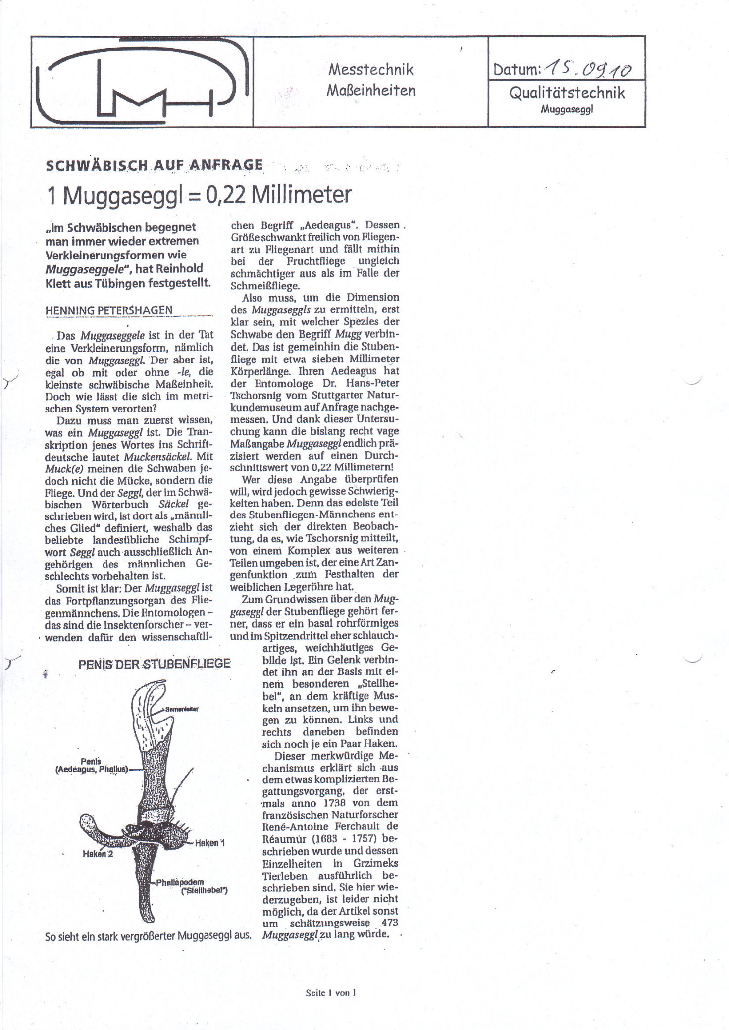
As muggeseggele ( houseflies a very small dimension in the Swabian dialect is -Aedeagus on Swabian) addressed. The similarly constructed English gnats cock (English gnat for mosquito ) is much more slang-like. According to measurements by an entomologist from the Stuttgart Natural History Museum, the aedeagus of a housefly (the Muggeseggele) is around 0.22 millimeters.
literature
- Wilfried Westheide , Reinhard Rieger (Hrsg.): Special zoology. Part 1: Protozoa and invertebrates. Gustav Fischer Verlag, Stuttgart et al. 1996, ISBN 3-437-20515-3 , pp. 650-651.
Individual evidence
- ↑ John L. Capinera (Ed.): Encyclopedia of Entomology. 2nd Edition. Springer, 2008, ISBN 978-1-4020-6242-1 , p. 7.
- ^ E. Mulsant: Notice sur Antoine-Casimir-Marguerite-Eugène Foudras. Préface Altisides. In: Histoire naturelle des coléoptères de France. Volume 13: Altisides. Term "Aedeagus", p. 32. (biodiversitylibrary.org)
- ↑ a b c S. von Kéler: Entomological dictionary with special consideration of the morphological terminology. 3. Edition. Akademie-Verlag, Berlin 1963.
- ↑ A. Peytoureau: Recherches sur l'anatomy et le développement de l'armure genital mâle of Lepidopteres. In: Comptes rendus hebdomadaires des séances de l'Académie des Sciences. Volume 118, January - June 1894, Paris 1894, p. 542. (biodiversitylibrary.org)
- ↑ A. Peytoureau: Contribution à l'étdude de la morphology de l'armure genital of Insectes. Paris 1895, p. 174. (biodiversitylibrary.org)
- ^ David Sharp: On the structure of the terminal segment in some male Hemiptera. In: Transactions of the Entomological Society of London for the year 1890. London 1890, p. 400. (biodiversitylibrary.org)
- ^ A b David Sharp, Frederic Muir: The comparative anatomy of the male genital tube in Coleoptera. In: Transactions of the Entomological Society of London for the year 1912. London 1912, p. 484 and p. 481 f. ( biodiversitylibrary.org )
- ↑ a b c d e f g R. E. Snodgrass: A revised interpretation of the external reproductive organs of male insects. In: Smithsonian miscellaneous collections. Vol. 135135, No. 6, Washington 1957, pp. 2 ff. (Repository.si.edu)
- ↑ a b Niels P. Kristensen: Lepidoptera, Moths and Butterflies. Vol. 2: Morphology, Physiology and Development. (= Handbook of Zoology. Volume IV, Part 36). de Gruyter, Berlin / New York 2003, ISBN 3-11-016210-5 , p. 103 in the Google book search
- ↑ Gerald William Krantz, David Evans Walter: A Manual of Acarology. Texas Tech University Press, 2009.
- ^ A b J. F. McAlpin et al: Manual of Nearctic Diptera. Volume 1, (= Research Branche, Agriculture Canada, Monograph. No. 27). Biosystematic Research Institute Ottawa, Ontario 1981, ISBN 0-660-10731-7 .
- ↑ a b Cedric Gillott: Entomology. 3. Edition. Springer, ISBN 1-4020-3182-3 .
- ↑ a b Malcolm Burr, K. Jordan: On Arixenia Burr, A Suborder of Dermaptera. In: 2nd International congress of entomology. Oxford, August 1912, Volume 1: Proceedings. P. 398 ff. ( Archive.org ).
- ^ Yoshitaka Kamimura: Right-handed penises of the earwig Labidura riparia (Insecta, Dermaptera, Labiduridae): Evolutionary relationships between structural and behavioral asymmetries . In: Journal of Morphology . tape 267 , no. 11 , 2006, ISSN 1097-4687 , p. 1381-1389 , doi : 10.1002 / jmor.10484 .
- ↑ Yoshitaka Kamimura: Possible Removal of Rival Sperm by the Elongated Genitalia of the Earwig, Euborellia plebeja . In: Zoological Society of Japan (Ed.): Zoological Science . tape 17 , no. 5 , 2000, ISSN 0289-0003 , p. 667-672 , doi : 10.2108 / zsj.17.667 .
- ↑ Karl J. Jarvis, Fabian Haas, Michael F. Whiting: Phylogeny of earwigs (Insecta: Dermaptera) based on molecular and morphological evidence: reconsidering the classification of Dermaptera . In: Systematic Entomology . tape 30 , no. 3 , 2005, ISSN 1365-3113 , p. 442-453 , doi : 10.1111 / j.1365-3113.2004.00276.x .
- ^ Fabian Haas: The phylogeny of the Forficulina, a suborder of the Dermaptera. In: Systematic Entomology. 20, 1995, pp. 85-98 ( bio-nica.info PDF).
- ↑ a b Richard Fox: Invertebrate Anatomy OnLine Periplaneta americana Copyright 2004 as html
- ↑ a b Heinz Freude , Karl Wilhelm Harde , Gustav Adolf Lohse (ed.): Die Käfer Mitteleuropas (= Käfer Mitteleuropas . Volume 1 : Introduction to Beetle Science ). 1st edition. Goecke & Evers, Krefeld 1965, ISBN 3-8274-0675-7 , pp. 26 .
- ↑ Heinz Freude, Karl Wilhelm Harde, Gustav Adolf Lohse (ed.): Die Käfer Mitteleuropas . tape 7 . Clavicornia. Spektrum Akademischer Verlag, Munich 1967, ISBN 3-8274-0681-1 , p. 280 .
- ↑ Chang S. Han, Piotr G. Jablonski: Female Genitalia Concealment Promotes Intimate Male Courtship in a Water Strider . In: PLoS ONE . tape 4 , no. 6 , 2009, ISSN 1932-6203 , doi : 10.1371 / journal.pone.0005793 , PMID 19516886 , PMC 2686155 (free full text).
- ↑ Representation of the partially everted genitals at ResearchGate.
- ↑ c: File: Melanoplus differentialis-Male-1.jpg and c: File: Melanoplus differentialis-Male-2.jpg by Eugene Zelenko
- ↑ of FIGS. 6 and 16.
- ↑ Aedeagus as an organ
- ↑ Aedeagus as a skeletal element
- ↑ giand.it
- ↑ c: File: Trichocera annulata, Trawscoed, North Wales, Dec 2016 2 - Flickr - janetgraham84.jpg
- ↑ Horst Neumann: The construction and function of the male genital apparatus of Trichocera annulata Meig. and Tipula paludosa Meig. In: German Entomological Journal. , NF 5, volume III / IV, 1958.
- ↑ c: File: Male genitalia Lepidoptera.jpg
- ↑ c: File: Neopalpa male genitalia.jpg
- ^ A b John R. Eyer: The comparitiv morphology of the male genitalia of the primitive Lepidoptera. Thesis, University of Minnesota, Dec. 1923, online as una-dissertation-0142.pdf
- ↑ A convex ring-shaped anellus in Digugua on the website Pictures of the reproductive system after the images of the imagines
- ↑ membranous, cap-shaped anellus
- ↑ Scheme of a funnel-shaped anellus in the third quarter of the page
- ↑ Manfred Koch , Wolfgang Heinicke, Bernd Müller: We determine butterflies. Volume 4: Spanner. 2nd, improved and enlarged edition. Neumann, Leipzig / Radebeul 1976, DNB 780451570 , p. 29.
- ↑ Designations according to Masao Ito: Supraspecific Classification of Bumblebees based on the Characters of Male Genitalia in Contributions from the Institute of Low Temperature Science, B. 20: 1 - 143 , Hokkaido University 1986-01-25 Doc. URL hdl.handle.net eprints.lib.hokudai.ac.jp
- ↑ Henning Petershagen: Swabian on request. 1 Muggaseggl = 0.22 millimeters . In: Data sheet measurement technology. Units of measurement → Quality technology: Muggaseggl from September 15, 2010; PDF, 497 kB; Retrieved July 7, 2013.