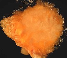
Granular cell tumor
| Classification according to ICD-O-3 | |
|---|---|
| 9580/0 | Granular cell tumor |
| 9580/3 | Malignant granular cell tumor |
| ICD-O-3 first revision online | |
The granular cell tumor (granular cell myoblastoma, myoblastic myoma, Abrikossow tumor) is a rare benign nonepithelial tumor, presumably of neuroectodermal origin, which usually manifests itself in middle age. The main locations are the tongue and the skin and subcutaneous tissue of the trunk. In addition, the tumor can occur in practically any anatomical location. Clinically, it is a slowly growing, usually painless tumor that rarely recurs after surgical removal . A malignant degeneration is observed in a small percentage of cases.
History
The granular cell tumor was first described in 1926 by Alexei Iwanowitsch Abrikossow , who initially interpreted the lesion as a benign neoplasm of the striated skeletal muscles and named it myoblastic myoma .
etiology
The underlying causes of the granular cell tumor are unknown. The Schwann cell is considered to be the likely starting point for tumor development , although a relationship cannot be established in all cases. The rare congenital variant of the gingival granular cell tumor may be a non- neoplastic , reactive lesion.
Epidemiology
The maximum age of the disease is in middle age, although the time of manifestation varies within wide limits. Women are affected slightly more often than men.
pathology
Macroscopically , they are mostly small, usually less than 3 cm in size, pale yellowish, often indistinctly delimited tumors of a solid consistency, which occur primarily in the tongue (40%) as well as in the skin and subcutaneous tissue, especially the trunk (30%). In addition, granular cell tumors have been described in many other locations, including the bronchial system (13%), urinary and genital apparatus (13%), gastrointestinal tract (6%), and the central nervous system .
Intracranially , the tumor occurs primarily in the pituitary region and is referred to here as a granular cell tumor of the neurohypophysis , which is classified as grade I according to the WHO classification of tumors of the central nervous system .
Histologically , granular cell tumors show a remarkably uniform appearance regardless of their location. The tumor cells are arranged in nests or cell balls, large, round, polygonal or elongated and have an abundance of fine-granular, eosinophilic cytoplasm in which larger eosinophilic droplets or granules are sometimes found. These contain plenty of hydrolytic enzymes such as acid phosphatase and can be regularly stained with the Luxol Fast Blue dye , in some cases also with the PAS stain. The cell boundaries are often indistinct, which can lead to the impression of a syncytial cell association. The cell nuclei are small, centrally located and mostly pyknotic or hyperchromatic , more rarely also vesicular. Mitoses and minor, often degenerative atypias are rarely observed. Often groups of tumor cells are found in the vicinity of small nerves. Tumors located on the surface are often accompanied by pseudoepitheliomatous hyperplasia of the covering squamous epithelium , which should not be confused with squamous cell carcinoma .
Immunohistochemistry
Immunohistochemically , the tumor cells of the granular cell tumor show a positivity for neuron-specific enolase (NSE), CD63 (NK1-C3), S-100 and almost always also for inhibin and calretinin . In addition, there is a fine-grained positivity for the lysosomal antigen CD68.
The rare malignant granular cell tumors often show negative immunoreactivity for NSE, S-100 and vimentin .
Diagnosis and differential diagnosis
The diagnosis is made after the pathologist has taken a tissue sample ( biopsy ) or on the preparation of the completely removed tumor . As a rule, the histological picture is so characteristic that diagnostic problems do not arise. Depending on the localization, the schwannoma , neurofibroma , alveolar soft tissue sarcoma, adult rhabdomyoma , histiocytoid carcinoma , leiomyoma or gastrointestinal stromal tumor and, rarely, reactive lesions following previous trauma or inflammation are possible differential diagnoses .
therapy
The treatment of choice is surgical removal of the tumor. A further safety margin to the tumor is only necessary for the malignant variant of the granular cell tumor.
forecast
As a usually benign neoplasm with slow growth, the granular cell tumor shows a good prognosis. The recurrence rate after surgical therapy is less than 5 percent; a recurrence of the tumor is usually due to incomplete removal. Malignant degeneration is observed in a maximum of 2–3 percent of cases. In the course of this, metastasis often occurs with an ultimately fatal outcome.
Individual evidence
- ↑ a b c d e f g h C. DM Fletcher: Diagnostic Histopathology of Tumors. 3. Edition. Churchill Livingstone, 2007.
- ↑ A. Abrikossoff: About myomas, starting from the striated voluntary muscles. In: Arch Pathol Anat. 1926; 260, p. 214.
- ^ A. Abrikossoff: Further investigations on myoblastic fibroids. In: Arch Pathol Anat. 1931; 280, p. 723.
- ↑ a b c d PathConsult: Granular Cell Tumor. (February 24, 2006), Elsevier; http://www.pathconsultddx.com/pathCon/diagnosis?TXTBOX2=gra&pii=S1559-8675%2806%2970247-9 ( page no longer available , search in web archives ) Info: The link was automatically marked as defective. Please check the link according to the instructions and then remove this notice.
- ↑ T. Schlick, T. Junginger: Abrikossoff granulosa cell tumor: a rare tumor of the esophagus. In: surgeon. 1997 Sep; 68 (9), pp. 932-935. PMID 9410685
- ↑ Cohen-Gadol et al .: Granular Cell Tumor of the Sellar and Suprasellar Region: Clinicopathologic Study of 11 Cases and Literature Review. In: Mayo Clin Proc. 2003; 78 (5), pp. 567-573. PMID 12744543 Full text ( page no longer available , search in web archives ) Info: The link was automatically marked as defective. Please check the link according to the instructions and then remove this notice.
- ^ A b The Maxillofacial Center for Education & Research: Granular Cell Tumor ; Archive link ( Memento of the original from January 26th, 2009 in the Internet Archive ) Info: The archive link has been inserted automatically and has not yet been checked. Please check the original and archive link according to the instructions and then remove this notice.


